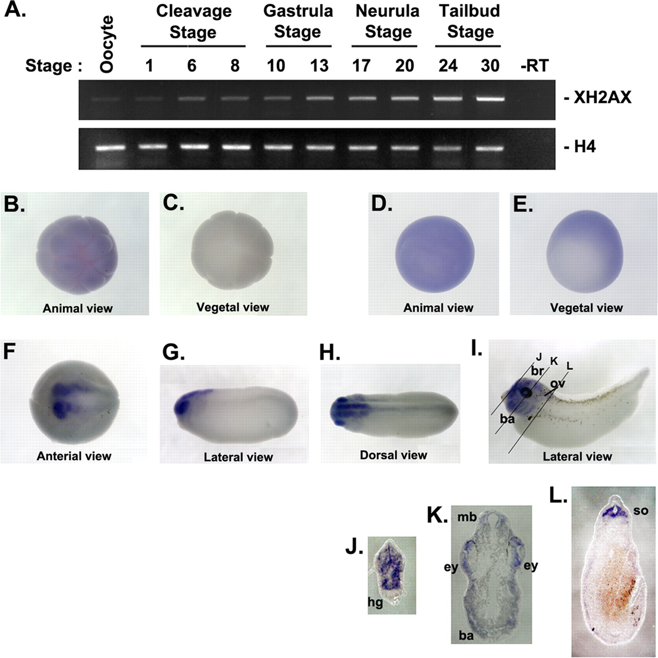
FIGURE 1. Temporal and spatial expression profile of XH2AX during embryogenesis. A, RT-PCR analysis of temporal mRNA expression of XH2AX. Developmental stages are indicated above each lane. Histone H4 served as a loading control. âRT, control reaction without reverse transcriptase. BâI, whole-mount in situ hybridization showing the spatial expression of XH2AX during early Xenopus development. B and C, blastula stage: animal hemisphere view (B) and vegetal hemisphere view (C); D and E, gastrula stage: animal hemisphere view (D) and vegetal hemisphere view (E); F, neurula stage: anterior view with posterior right; G and H, tail bud stage: lateral view with anterior left (G) and dorsal view with anterior left (H); I, stages 33â34. The black lines represent the angle of sectioning for JâL. JâL, transverse section through a stage 33â34 embryo stained by whole-mount in situ hybridization for XH2AX mRNA. ba, branchial arches; br, brain; ey, eye; hg, hatching gland; mb, midbrain; ov, otic vesicle; so, somite.
Image published in: Lee SY et al. (2010)
Copyright © 2010. Image reproduced with permission of the Publisher and the copyright holder. This is an Open Access article distributed under the terms of the Creative Commons Attribution License.
| Gene | Synonyms | Species | Stage(s) | Tissue |
|---|---|---|---|---|
| h2ax.S | h2afx, h2ax.1, XH2AX | X. laevis | Sometime during NF stage 7 to NF stage 33 and 34 | animal cap eye eye primordium optic vesicle brain forebrain midbrain hindbrain animal hemisphere |
Image source: Published
Permanent Image Page
Printer Friendly View
XB-IMG-134535