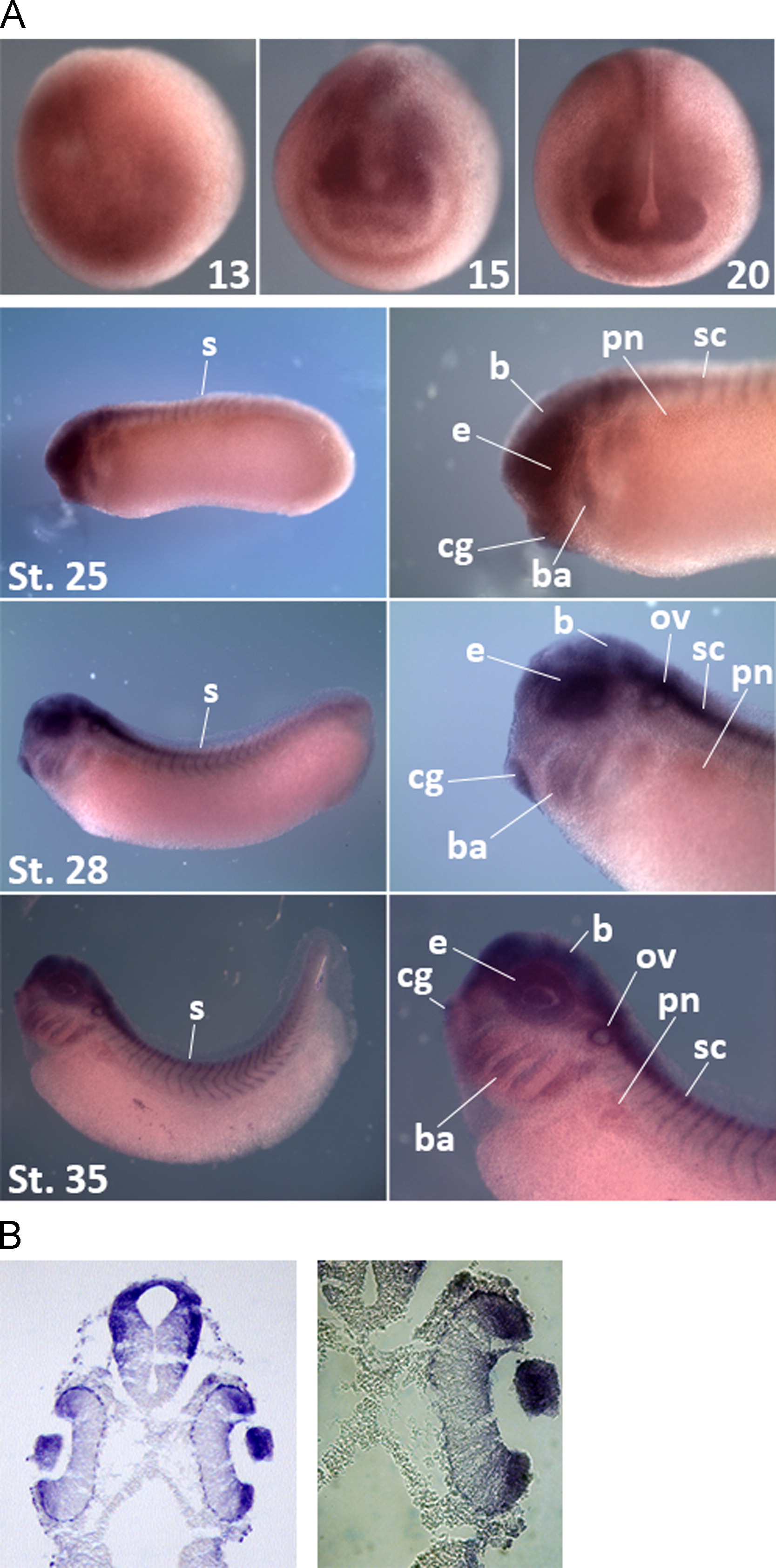
Fig. 2. Spatial expression patterns of XFrs3. (A) Whole-mount in situ hybridization of XFrs3. Embryos at indicated stages (13, 15, 20, 25, 28 and 35) were subjected to in situ hybridization with XFrs3 antisense probe. For stages 25, 28 and 35, right panels show magnified images of embryos at left panels. At early neurula stage (stage 13), XFrs3 is expressed broadly in the presumptive anterior neuroectoderm, and its expression is gradually localized to the eye field, cement gland and neural tube at mid-neurula and late neurula stages (stages 15 and 20). At early tailbud stages (stages 25 and 28) and tadpole stage (stage 35), XFrs3 transcript is localized in anterior structures of neuroectodermal origin [branchial arches (ba), cement gland (cg), eyes (e) and otic vesicles (ov)], brain (b), spinal cord (sc), somites (s) and pronephros (pn). (B) XFrs3 expression pattern in the eye at stage 35. Embryos at stages 35 were subjected to whole-mount in situ hybridization with XFrs3 antisense probe and then cross-sectioned. XFrs3 is expressed in the lens, the retina, and the brain.
Image published in: Kim YJ et al. (2015)
Copyright © 2015. Image reproduced with permission of the Publisher, Elsevier B. V.
| Gene | Synonyms | Species | Stage(s) | Tissue |
|---|---|---|---|---|
| frs3.S | X. laevis | Throughout NF stage 13 | neuroectoderm anterior ectoderm | |
| frs3.S | X. laevis | Sometime during NF stage 15 to NF stage 20 | neural tube anterior neural fold eye primordium cement gland primordium neural crest | |
| frs3.S | X. laevis | Sometime during NF stage 25 to NF stage 35 and 36 | lens eye retina brain spinal cord cement gland neural tube somite pharyngeal arch otic vesicle pronephric mesenchyme |
Image source: Published
Permanent Image Page
Printer Friendly View
XB-IMG-134789