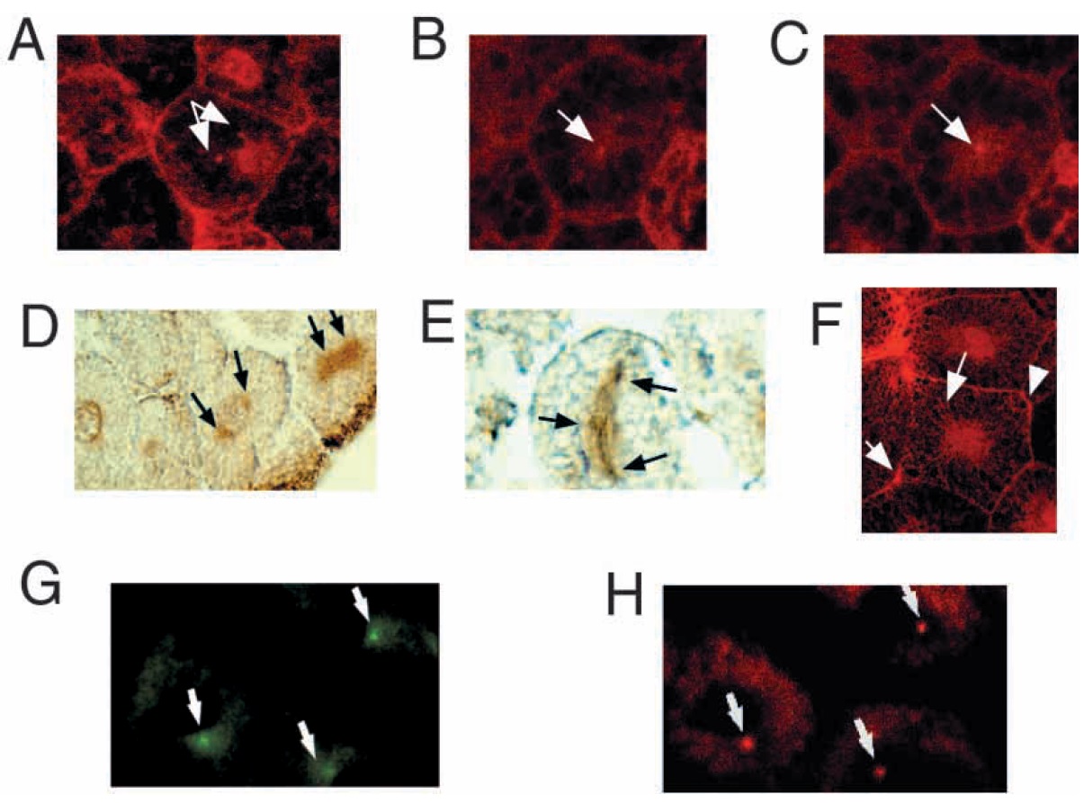
Fig. 3. Intracellular distribution of endogenous and exogenous XCS-1 protein. (A-C) Confocal images of whole mounts of stage 10 embryos immunostained using the affinity purified anti-XCS-1 antibody to detect endogenous XCS-1. (A) Arrows are pointing to the XCS-1 protein located on two structures that appear to be centrosomes; (B and C) different focal planes through the same blastomere showing centrosomes on different sides of the nucleus; (D,E,H) in vitro-transcribed RNA from myc-XCS-1 (10 pg) was injected into fertilized eggs. Stage 10 embryos were fixed and sectioned and immunostained with anti-myc antibody or anti-g-tubulin antibody. (D and E) Peroxidase-staining using anti-myc antibody â arrows are pointing to the centrosomal area, the spindle and to the chromosomes on the spindle. (F) Confocal image of endogenous XCS-1 protein decorating intracellular structures as well as cell membrane; (G) indirect immunofluorescence with the anti myc-XCS-1 antibody showing intracellular distribution of exogenous XCS-1 at the centrosomal area of 3 cells (arrows). (H) The same cells in panel G that were immunostained with anti g-tubulin antibodies to show the position of the centrosomes (arrows).
Image published in: Nakamura H et al. (2000)
Copyright © 2000. Image reproduced with permission of the Publisher, The Company of Biologists Ltd.
| Gene | Synonyms | Species | Stage(s) | Tissue |
|---|---|---|---|---|
| itprid2.S | ssfa2, XCS-1 | X. laevis | Throughout NF stage 10 | centrosome mitotic cell |
| tubg1.L | gamma tubulin, gamma-tubulin, Xgam | X. laevis | Throughout NF stage 10 | cell centrosome |
Image source: Published
Permanent Image Page
Printer Friendly View
XB-IMG-135174