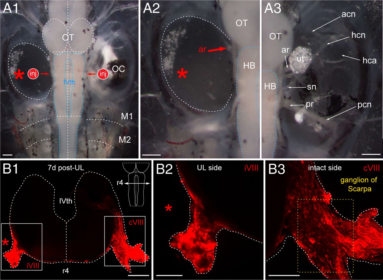
Figure 6.nMorphological consequences of UL on VIIIth nerve afferent fibers in a stage 55 Xenopus tadpole. A, Dorsal view of the brainstem/spinal cord and bilateral otic capsules (A1) 7 d after a UL on the left side (red asterisk) along with an indication of the sites of fluorescent tracer application (inj) to the severed, left and intact, right VIIIth nerve; higher magnification of the otic capsules on both sides illustrate the absence of all labyrinthine end organs on the operated (A2) and their presence on the intact side (A3). B, Confocal reconstruction of a cross-section through the hindbrain at r4 (schematic inset in B1) depicting vestibular nerve afferent fibers on both sides at the entrance of the anterior branch (ar) of the VIIIth nerve into the brainstem 7 d postlesion; fibers were labeled after tracer application to the nerve stump on the operated side and the respective location on the intact side (A1, inj); higher magnification of the outlined areas in B1 illustrating the reduction of labeled fibers on the operated (B2) with respect to the intact (B3) side. HB indicates hindbrain; OT, optic tectum; M1â2, myotome 1â2; IVth, IVth ventricle; ar and pr, anterior and posterior branch of the VIIIth nerve; acn, pcn, and hcn, anterior, posterior, and horizontal semicircular canal nerve; hca, horizontal semicircular canal ampulla; sn, saccular nerve; ut, utricle; iVIII and cVIII, ipsilesional and contralesional VIIIth nerve. Scale bar in A1âA3 represents 300 μm; in B1, 200 μm; and in B2, B3, 100 μm, respectively.
Image published in: Lambert FM et al. (2013)
Copyright © 2013. OA ARTICLE, images redisplayed under a Creative Commons license.
Permanent Image Page
Printer Friendly View
XB-IMG-138220