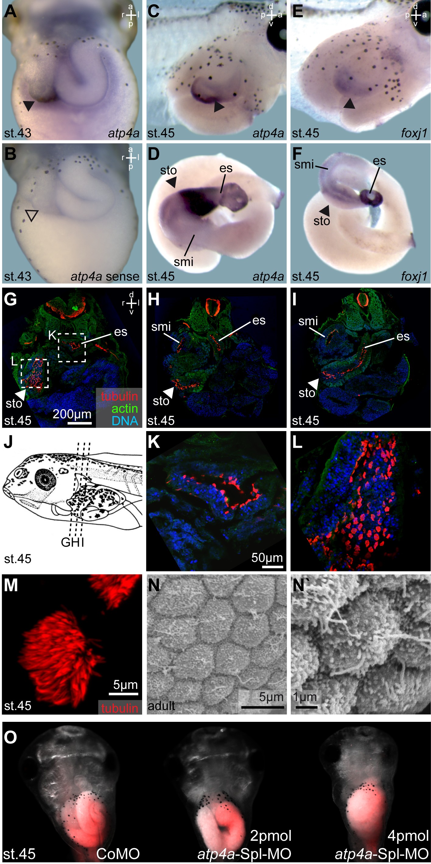
Fig. 5. foxj1 and atp4a are co-expressed in the gastrointestinal tract which transiently harbors motile cilia in the stomach. (AâF) WMISH for atp4a (A,C,D) and foxj1 (E,F) in the GI tract of stage 43â45 tadpoles (AâC, E; stomach highlighted by arrowheads; ventral views) and isolated GI tracts (D, F). (B) atp4a sense control revealed no staining. Note the co-expression of atp4a and foxj1 at stage 45 (CâF). (GâM) GI tract ciliation as shown by immunofluorescent staining of cilia/tubulin (acetylated-α-tubulin staining, red) and staining for actin (phalloidin, green) as well as nuclei (DAPI, blue) on cryosections from stage 45 tadpoles (planes indicated in J). MCCs (M) were found in the esophagus (es; GâI,K), stomach (sto; GâI,L) and the proximal part of the small intestine (smi; H, I). (N, N`) Scanning electron microscopy analysis of gastric epithelia from adult frogs revealed the presence of short monocilia, but lack of MCCs. (O) Injection of atp4aSplMO targeted to the endoderm prevented normal development of the GI tract. Targeting was monitored by co-injection of fluorescent rhodamine dextrane (red). Embryos are shown in ventral view. a, anterior; d, dorsal; es, esophagus; l, left; p, posterior; r, right; st., stage; smi, small intestine; sto, stomach; v, ventral.
Image published in: Walentek P et al. (2015)
© 2015 The Authors. Creative Commons Attribution license
| Gene | Synonyms | Species | Stage(s) | Tissue |
|---|---|---|---|---|
| atp4a.L | alpha H-K, gastric H(+)-K(+)-ATPase alpha-subunit, LOC108696872, MGC69393 | X. laevis | Sometime during NF stage 43 to NF stage 45 | alimentary system esophagus stomach intestine ciliated cell |
| tuba4b.L | alpha tubulin, alpha-tubulin, tuba4, tuba4a | X. laevis | Throughout NF stage 45 | esophagus stomach small intestine multiciliated cell ciliated cell alimentary system |
| foxj1.L | foxj1.1, foxj1-a, foxj1-b, hfh-4, hfh4, xfoxj1 | X. laevis | Throughout NF stage 45 | esophagus stomach intestine small intestine alimentary system |
Image source: Published
Permanent Image Page
Printer Friendly View
XB-IMG-144173