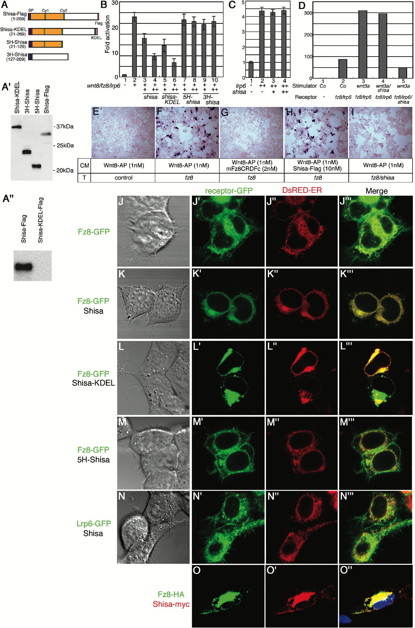
Figure 4. Shisa Cell-Autonomously Inhibits Wnt Signaling by Retaining Fz within the ER (A) Schematic drawing of Shisa, fused with the ER retention KDEL signal, and deletion constructs employed. Shisa-KDEL, 5H-Shisa, and 3H-Shisa were generated by fusing a cassette containing a heterologous signal peptide followed by a Flag sequence to shisa cDNA fragments. Numbers in parentheses indicate corresponding Shisa amino acid residues. (Aâ²) Western blotting with α-Flag mAb shows equivalent protein productions of each construct. Cells were transfected with each construct (100 ng) in 12-well plate. (Aâ²) KDEL signal suppressed secretion of Shisa. Cells were transfected as in (Aâ²). (B) Shisa and Shisa-KDEL, but not 5H- and 3H-shisa, inhibited TOPFLASH reporter activation induced by XWnt8-Fz8-Lrp6 expression. Luciferase activities are indicated as fold activation/repression compared with the activity obtained from cells transfected with empty-vector and reporter (lane 1). Each experiment was carried out at least in triplicate, and error bars represent the standard deviation. Transfection was carried out in a 96-well plate with DNAs (per well): TOPFLASH reporter, 10 ng; nlacZ, 1 ng; Xwnt8, 5 ng; fz8, 1 ng; lrp6, 1 ng; shisa, shisa-KDEL, 5H-shisa, and 3H-shisa, 5 ng (+) or 25 ng (++). (C) Shisa failed to inhibit the signaling induced by a high dose of Lrp6 alone. DNA used: lrp6, 20 ng; shisa 5 ng for lane 3; and 25 ng for lane 4. (D) Shisa cell-autonomously inhibited Wnt signaling in the cells receiving the signal. Stimulator and receptor cells were transfected separately and mixed in the combination presented at the bottom of the figure. Co: Cells transfected with the empty-vector alone. DNA used: mwnt3a, 500 ng; fz8, 8 ng; lrp6, 8 ng; TOPFLASH reporter, 80 ng; nlacZ, 8 ng; shisa, 200 ng. (EâI) Shisa suppressed Wnt8-AP and Fz8 interaction. Live cells were stained with 1 nM of Wnt8-AP. Cells in an 8-well chamber slide were transiently transfected with DNAs: fz8, 2 ng; shisa, 50 ng. CM, condition media used. T, construct used for transfection. (JâOâ²) Confocal immunofluorescent images of HEK 293T cells. Phase contrast images were (J), (K), (L), (M), and (N). ER was marked by DsRedER in (Jâ²), (Kâ²), (Lâ²), (Mâ²), and (Nâ²). (JâJâ´) Transfected with fz8-GFP (green) (cell surface expression of Fz, n = 100, 98%). (KâKâ´) Transfected with fz8-GFP and shisa (ER retention of Fz, n = 100, 90%). (LâLâ´) Transfected with fz8-GFP and shisa-KDEL (ER retention of Fz, n = 100, 84%). (MâMâ´) Transfected with fz8-GFP and 5H-shisa (cell surface expression of Fz, n = 100, 92%). (NâNâ´) Transfected with lrp6-GFP and shisa (cell surface expression of Lrp6, n = 100, 86%). (OâOâ²) Transfected with fz8-HA and shisa-myc (colocalization, n = 100, 84%). Cells were transfected in an 8-well glass chamber with DNAs: fz8-GFP, 2 ng; Lrp6-GFP, 2 ng; fz8-HA, 2 ng; shisa, 50 ng; shisa-KDEL, 50 ng; 5H-shisa, 50 ng; shisa-Myc, 50 ng; pDsRed-ER, 10 ng.
Image published in: Yamamoto A et al. (2005)
Copyright © 2005. Image reproduced with permission of the Publisher, Elsevier B. V.
Permanent Image Page
Printer Friendly View
XB-IMG-145789