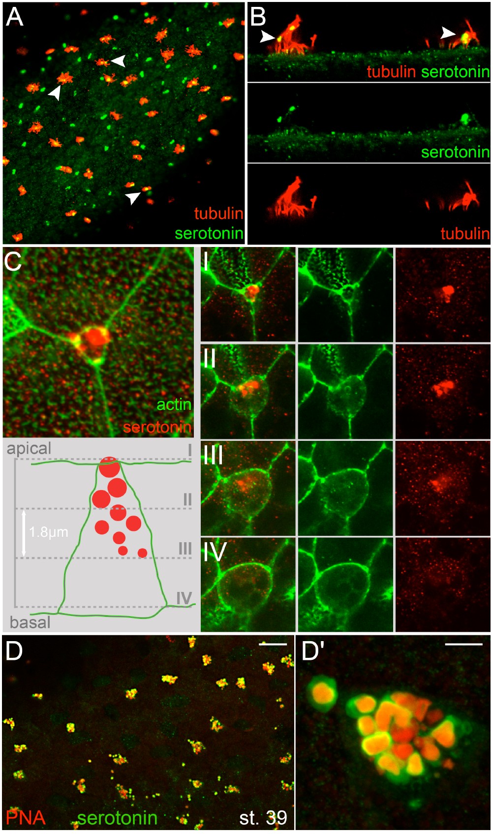
Fig. S1. Cilia association of serotonin-containing vesicles and vesicle maturation. (A, B) Co-staining of serotonin (green) and cilia (red; anti acetylated-ô-tubulin) at stage 32 reveals occasional co-localization in the epidermis (white arrowheads). (B) Higher magnification of two MCCs in side view. Note the vesicular nature of serotonin staining in all cases. (C) Vesicle maturation in SSCs. Z-stack analysis of a single SSC stained for serotonin (red) and actin (phalloidin, green). Apical to basal horizontal optical sections (I-V) were used to reconstruct an orthogonal hypothetical model of vesicle maturation (lower panel in C). Vesicles localized basally were small (0.2 μm diameter), with a continuous increase in diameter to a maximum of 1 μm towards the apical pole of the cell. (D, D') Vesicular co-localization of serotonin (green) and peanut agglutinin (PNA; red) in the epidermis of Xenopus laevis tadpoles at stage 39. Scale bars in (D) and (D') represent 50 and 5μm, respectively.
Image published in: Walentek P et al. (2014)
Copyright © 2014. Image reproduced with permission of the Publisher.
| Gene | Synonyms | Species | Stage(s) | Tissue |
|---|---|---|---|---|
| tuba4a.L | alpha tubulin, alpha-tubulin, tuba4, tuba4b | X. laevis | Throughout NF stage 32 | epidermis epithelium cilium ciliated cell mucociliary epithelium |
Image source: Published
Permanent Image Page
Printer Friendly View
XB-IMG-151069