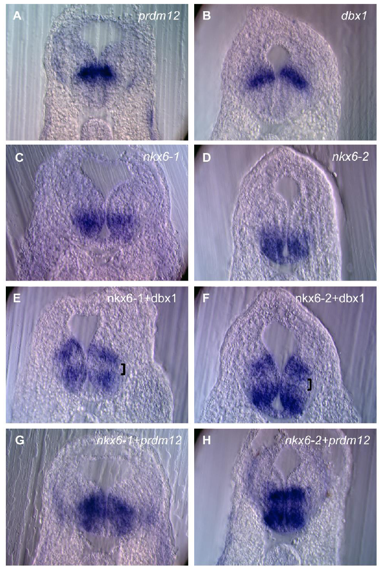
Fig. S5. prdm12 expression compared to dbx1, nkx6-1 and nkx6-2 in the posterior hindbrain of Xenopus tailbud stage embryos. (A-H) Transverse sections of the neural tube of stage 28 embryos hybridized with the indicated probes. In the double in situ hybridization experiments shown in E-H, both probes are revealed in dark blue. Note the gap of staining between nkx6-1 or nkx6-2 and dbx1, which corresponds to the p1 progenitor domain (brackets in E, F) and the absence of this gap between nkx6-1 or nkx6-2 and prdm12.
Image published in: Thélie A et al. (2015)
Copyright © 2015. Image reproduced with permission of the Publisher and the copyright holder. This is an Open Access article distributed under the terms of the Creative Commons Attribution License.
| Gene | Synonyms | Species | Stage(s) | Tissue |
|---|---|---|---|---|
| prdm12.L | LOC108699953, pfm9 | X. laevis | Throughout NF stage 28 | hindbrain |
| dbx1.S | dbx, Xdbx | X. laevis | Throughout NF stage 28 | hindbrain |
| nkx6-1.L | LOC108704117, nkx6.1 | X. laevis | Throughout NF stage 28 | hindbrain |
| nkx6-2.L | nkx6.2 | X. laevis | Throughout NF stage 28 | hindbrain |
Image source: Published
Permanent Image Page
Printer Friendly View
XB-IMG-151842