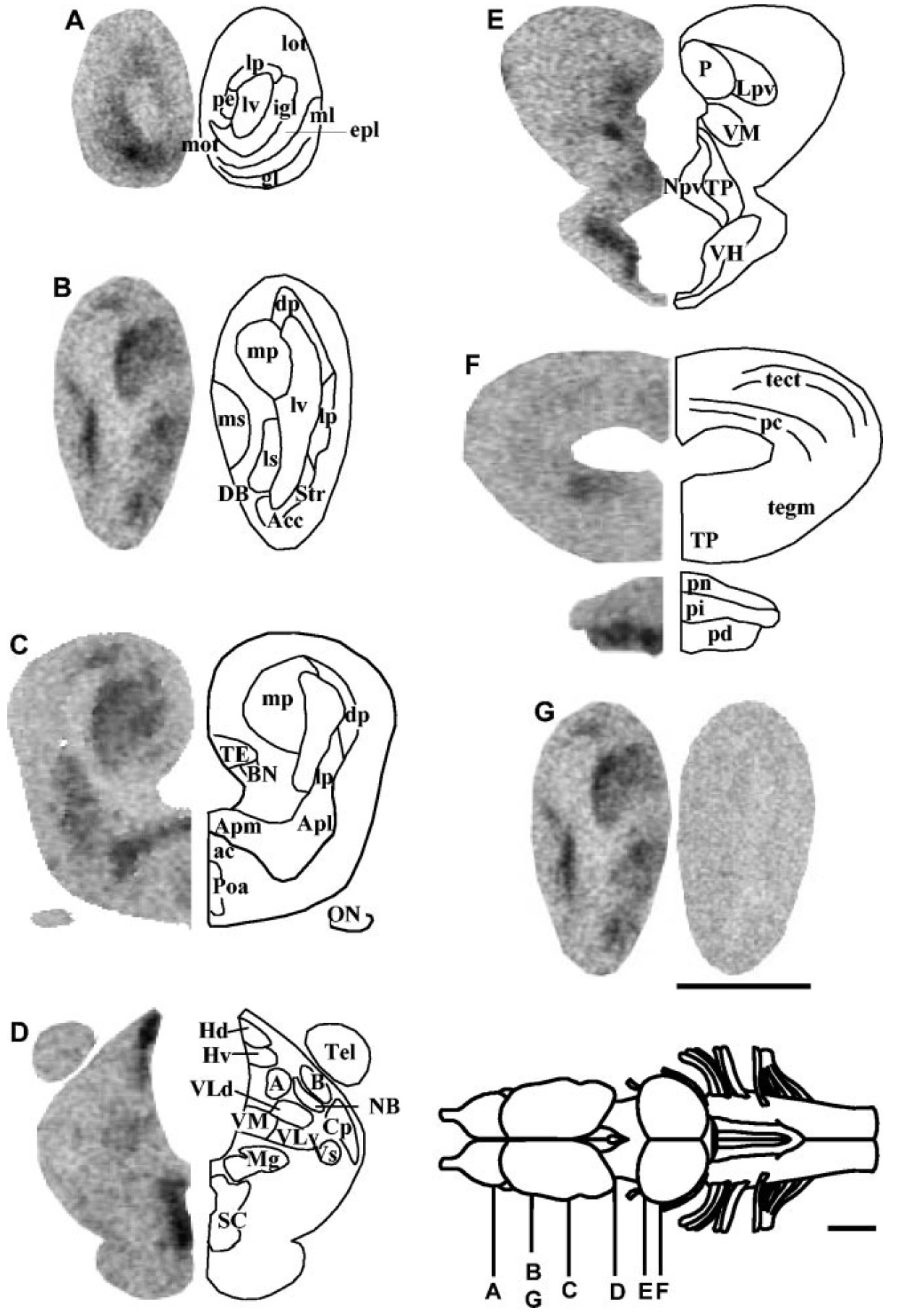
Fig. 5. In situ hybridization histochemistry showing the distribution of xTRHR2 mRNA in the brain and pituitary of white-adapted Xenopus laevis. AâF: Frontal brain sections from white-adapted frogs were hybridized with an antisense xTRHR2 receptor riboprobe and exposed onto Hyperfilm max for 2 weeks. G: A control section incubated with a sense riboprobe (right hemisection) is compared with a consecutive section hybridized with the antisense probe (left hemisection). See legend to Figure 1 for other designations. Scale bars 1 mm.
Image published in: Bidaud I et al. (2004)
Copyright © 2004. Image reproduced with permission of the Publisher.
| Gene | Synonyms | Species | Stage(s) | Tissue |
|---|---|---|---|---|
| trhr2.L | LOC108704068, xtrhr2 | X. laevis | Throughout NF stage 66 | brain hypophysis suprachiasmatic nucleus habenula dorsal habenular nucleus amygdala lateral amygdala medial amygdala medial pallium medial septum thalamus ventromedial thalamic nucleus granule cell layer of the olfactory bulb |
Image source: Published
Permanent Image Page
Printer Friendly View
XB-IMG-151903