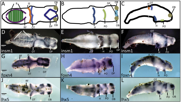
Fig. 2. insm1 expression in the developing brain. A â C. Diagrammatic representation of selected brain regions as viewed in dorsally (A), ventrally (B) or laterally (C). D â L. Whole mount in situ hybridizations of stage 45 dissected brains using probes for insm1 (D â F), foxn4 (G â I), or lhx5 (J â L). insm1 is expressed in the dorsal pallium, optic tectum and rhombomeres as viewed dorsally (D). insm1 expression was also detected ventrally (E) and laterally (F) in the hypothalamus, preoptic area and isthmus. Scale bars (1 μM) are indicated in (D) for (D â F), (G) for (G â I) and (J) for (J â L). Abbreviations: DP â dorsal pallium, H â hypothalamus, IS â isthmus, OB â olfactory bulb, OT â optic tectum, PO â preoptic area, RF â reticular formation, R â rhombomeres.
Image published in: Bosse JL and El-Hodiri HM (2016)
Copyright © 2016. Image reproduced with permission of the Publisher, Elsevier B. V.
| Gene | Synonyms | Species | Stage(s) | Tissue |
|---|---|---|---|---|
| insm1.L | IA1 | X. laevis | Throughout NF stage 45 | brain hindbrain dorsal pallium optic tectum hypothalamus preoptic area |
| foxn4.L | X. laevis | Throughout NF stage 45 | brain hindbrain midbrain-hindbrain boundary optic tectum dorsal pallium hypothalamus preoptic area | |
| lhx5.L | xlim-2a, xlim-5, Xlim5 | X. laevis | Throughout NF stage 45 | brain hypothalamus preoptic area olfactory bulb hindbrain midbrain forebrain |
Image source: Published
Permanent Image Page
Printer Friendly View
XB-IMG-153537