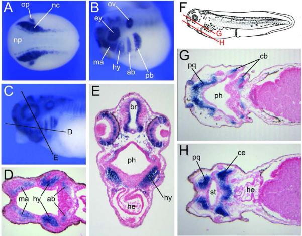
Figure 2. Developmental expression of Xenopus Sox9 in the neural crest lineage (A) By in situ hybridization, at the end of gastrulation Sox9 is detected in neural crest progenitors (nc) at the lateral edge of the neural plate (np), and in the presumptive otic placode (op). Dorsal view, anterior to left. (B) At the tailbud stage Sox9 is detected in the four streams of cranial neural crest migrating towards the pharyngeal arches, the mandibular (ma), hyoid (hy), anterior branchial (ab) and posterior branchial (pb) neural crest. Other domains of expression include the developing eye (ey) and the otic vesicle (ov). Lateral view, anterior to left, dorsal to top. (C) Sox9 expression in the head region of a stage 35 embryo (Nieuwkoop and Faber, 1967). Lateral view, anterior to left, dorsal to top. The black lines indicate the level of the sections shown in the subsequent panels. (DâE) Sections showing Sox9 expression in the mesenchyme of the pharyngeal arches. (F) Diagram of a stage 40 embryo, after Nieuwkoop and Faber (1967). Lateral view, anterior to left, dorsal to top. The red lines indicate the level of the sections shown in the subsequent panels. (GâH) Sox9 is detected in all differentiating cartilage elements, including the palatoquadrate (pq), ceratobranchial (cb), ceratohyal (ce) and Meckel's cartilage (not shown). br, brain; he, heart; ph, pharynx; st, future stomodeum.
Image published in: Lee YH and Saint-Jeannet JP (2011)
Copyright © 2011. Image reproduced with permission of the Publisher.
| Gene | Synonyms | Species | Stage(s) | Tissue |
|---|---|---|---|---|
| sox9.L | cmd1, cmpd1, sox-9, sox9-a, sox9-b, sra1 | X. laevis | Sometime during NF stage 17 to NF stage 18 | neural plate border neural crest premigratory neural crest cell otic placode neuroectoderm |
| sox9.L | cmd1, cmpd1, sox-9, sox9-a, sox9-b, sra1 | X. laevis | Throughout NF stage 28 | otic vesicle brain eye cranial neural crest mandibular crest hyoid crest anterior branchial crest posterior branchial crest |
| sox9.L | cmd1, cmpd1, sox-9, sox9-a, sox9-b, sra1 | X. laevis | Throughout NF stage 35 and 36 | pharyngeal mesenchyme pharyngeal arch mandibular arch hyoid arch branchial arch cartilage element palatoquadrate ceratobranchial I ceratohyal Meckel's cartilage ceratobranchial II ceratobranchial III ceratobranchial IV |
Image source: Published
Permanent Image Page
Printer Friendly View
XB-IMG-153733