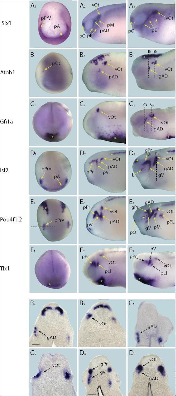
Figure 4âfigure supplement 4. Expression of targets with persistent expression in placodes in whole-mount Xenopus embryos. Expression dynamics for each target are shown across a range of developmental stages: A1âF1 show expression in neural plate stage embryos, A2âF2 show early tail bud stage embryos and A3âF3 show late tail bud stage embryos. B4âD4 and B5 âD5 shows sections at level indicated in B3âD3, respectively (dotted lines). (A) Expression of PPE marker gene Six1 is shown as reference for placodal domains (for details see Pandur and Moody, 2000; Schlosser and Ahrens, 2004). (B) Atoh1 is initially expressed at very low levels in presumptive otic placodes at neural plate stages (B1). This expression becomes more pronounced in the otic vesicle at early tail bud stages (B2) concomitant with the initiation of expression in lateral line ganglia and strong expression in the hindbrain (B2; asterisk). Expression becomes more pronounced in all three regions at late tail bud stages (B3 âB5). (C) Gfi1a is expressed at high levels in haematopoietic cells during neural plate stages (C1; asterisk). At early tail bud stages (C2) expression becomes more pronounced and diffuse, and expression is also initiated in the otic vesicle. At late tail bud stages Gfi1a is expressed in lateral line placodes as well as otic vesicles as the haematopoietic expression begins to subside (C3âC5). (D) During neural plate stages Isl2 is expressed in the profundal and trigeminal placodes and in the anterior placodal region along the anterior edge of the neural plate (D1). At early tail bud stages Isl2 expression is maintained in the profundal and trigeminal placodes/ganglia as well as in otic and lateral line placodes/ganglia and primary neurons in the spinal cord (D2; asterisk). Expression is maintained in cranial ganglia at late tail bud stages (D3âD5) and becomes apparent in the forebrain and lens (D3; arrowhead). (E) During neural plate stages Pou4f1.2 is expressed in the profundal and trigeminal placodes as well as in a stripe of primary sensory neurons (E1; asterisk; see Figure 4âfigure supplement 5 for section). In early tail bud stages (E2) expression in the profundal/trigeminal placodes/ganglia and primary neurons is maintained, and expression in the otic and lateral line placodes is strengthened. Expression is maintained in all domains as well as in the cranial ganglia derived from placodes into late tail bud stages when expression becomes up-regulated in the retina (E3; diamond). Dotted line in E1 indicates the level of section shown in Figure 4â figure supplement 5. (F) Tlx1 is expressed in the presumptive ventral visceral arches at neural plate stages (F1; asterisk). This is maintained into early and late tail bud stages (F2 and F3), which also exhibit prominent expression in the profundal/trigeminal placodes and ganglia and the otic vesicle. Yellow and black arrows mark placodal expression. Bar in B4, C4 and D4: 100 μm (also for B5, C5 and D5, respectively). Abbreviations: pA: anterior placodal region; pAD: anterior lateral line placode; gAD: ganglion of the anterodorsal lateral line nerve; pE: epibranchial placode; pLl: lateral line placodes; L: lens; pL: lens placode; pM: middle lateral line placode; pO: olfactory placode; vOt: otic vesicle; pOt: presumptive otic placode; pPr: profundal placode; pP: posterior placodal region; pPL: posterior lateral line placode; pPrV: profundal/trigeminal placodes; pV: trigeminal placode; gV: ganglion of the trigeminal nerve.
Image published in: Riddiford N and Schlosser G (2016)
© 2016, Riddiford et al. Creative Commons Attribution license
| Gene | Synonyms | Species | Stage(s) | Tissue |
|---|---|---|---|---|
| six1.L | XSix1 | X. laevis | Throughout NF stage 17 | profundal placode trigeminal placode anterior placodal area |
| atoh1.L | ath1, bhlha14, hath1, math-1, math1, xath-1 | X. laevis | Throughout NF stage 17 | olfactory placode |
| isl2.L | islet-2, islet2b, LOC108711275 | X. laevis | Throughout NF stage 17 | profundal placode trigeminal placode anterior placodal area |
| pou4f4.L | Brn3d, pou4f1.2 | X. laevis | Throughout NF stage 17 | profundal placode trigeminal placode neuron |
| gfi1.L | gfi1a | X. laevis | Throughout NF stage 18 | ventral blood island hematopoietic stem cell anterior ventral blood island ventral mesoderm |
| tlx1.L | hox11, hox11-a, LOC108697427, Xhox11, XHox11L2 | X. laevis | Throughout NF stage 18 | ventral mesoderm |
| six1.L | XSix1 | X. laevis | Throughout NF stage 24 | lateral line placode otic vesicle otic placode lateral line ganglion posterior lateral line ganglion olfactory placode epibranchial placode facial epibranchial placode glossopharyngeal epibranchial placode |
| atoh1.L | ath1, bhlha14, hath1, math-1, math1, xath-1 | X. laevis | Throughout NF stage 24 | lateral line placode otic placode otic vesicle hindbrain pharyngeal mesenchyme |
| gfi1.L | gfi1a | X. laevis | Throughout NF stage 24 | otic placode ventral blood island hematopoietic stem cell anterior ventral blood island |
| isl2.L | islet-2, islet2b, LOC108711275 | X. laevis | Throughout NF stage 24 | lateral line placode profundal placode trigeminal placode otic placode spinal cord neuron |
| pou4f4.L | Brn3d, pou4f1.2 | X. laevis | Throughout NF stage 24 | lateral line placode profundal placode trigeminal placode otic placode spinal cord spinal neuron |
| tlx1.L | hox11, hox11-a, LOC108697427, Xhox11, XHox11L2 | X. laevis | Throughout NF stage 24 | lateral line placode profundal placode otic placode ventral mesoderm |
| six1.L | XSix1 | X. laevis | Throughout NF stage 28 | otic vesicle olfactory placode epibranchial placode facial epibranchial placode glossopharyngeal epibranchial placode vagal epibranchial placode |
| atoh1.L | ath1, bhlha14, hath1, math-1, math1, xath-1 | X. laevis | Throughout NF stage 28 | lateral line placode otic vesicle hindbrain |
| gfi1.L | gfi1a | X. laevis | Throughout NF stage 28 | lateral line placode otic vesicle posterior ventral blood island ventral blood island |
| isl2.L | islet-2, islet2b, LOC108711275 | X. laevis | Throughout NF stage 28 | lateral line placode lens placode forebrain profundal placode profundus ganglion trigeminal placode trigeminal ganglion otic vesicle lateral line ganglion spinal cord neuron |
| pou4f4.L | Brn3d, pou4f1.2 | X. laevis | Throughout NF stage 28 | lateral line placode profundus ganglion trigeminal ganglion lateral line ganglion posterior lateral line ganglion otic vesicle olfactory placode |
| tlx1.L | hox11, hox11-a, LOC108697427, Xhox11, XHox11L2 | X. laevis | Throughout NF stage 28 | profundal placode trigeminal placode trigeminal ganglion otic vesicle ventral mesoderm |
Image source: Published
Permanent Image Page
Printer Friendly View
XB-IMG-153798