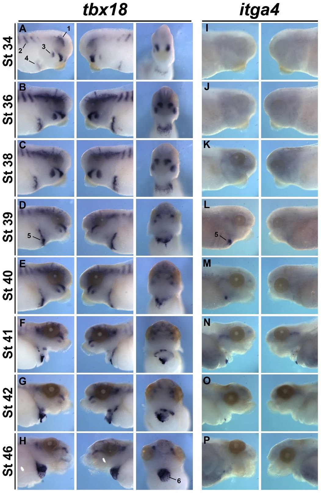
Figure S3. Spatio-temporal analysis of tbx18 and itga4 during Xenopus embryogenesis. (A-H) In situ hybridization of tbx18 showing right, left and ventral views of wild-type embryos from stage 34 to 46, anterior region of embryo. (I-P) In situ hybridization of itga4 showing right and left views of anterior region from stage 34-46. 1; cranial mesoderm, 2; somites, 3; branchial arches, 4; septum transversum, 5; proepicardial cluster, 6; epicardium.
Image published in: Tandon P et al. (2016)
Copyright © 2016. Image reproduced with permission of the Publisher and the copyright holder. This is an Open Access article distributed under the terms of the Creative Commons Attribution License.
| Gene | Synonyms | Species | Stage(s) | Tissue |
|---|---|---|---|---|
| tbx18.L | X. laevis | Throughout NF stage 33 and 34 | mesoderm head mesoderm axial mesoderm pharyngeal region proepicardium head | |
| tbx18.L | X. laevis | Throughout NF stage 35 and 36 | head head mesoderm axial mesoderm proepicardium pharyngeal region epicardium heart | |
| tbx18.L | X. laevis | Throughout NF stage 37 and 38 | head pharyngeal region somite proepicardium epicardium heart | |
| tbx18.L | X. laevis | Throughout NF stage 39 | head somite proepicardium epicardium heart | |
| itga4.L | inta4 | X. laevis | Throughout NF stage 39 | proepicardium |
| tbx18.L | X. laevis | Throughout NF stage 40 | head somite proepicardium epicardium heart | |
| itga4.L | inta4 | X. laevis | Throughout NF stage 40 | proepicardium head |
| tbx18.L | X. laevis | Throughout NF stage 41 | head proepicardium epicardium heart | |
| itga4.L | inta4 | X. laevis | Throughout NF stage 41 | head proepicardium |
| tbx18.L | X. laevis | Throughout NF stage 42 | head proepicardium epicardium heart | |
| itga4.L | inta4 | X. laevis | Throughout NF stage 42 | proepicardium |
| tbx18.L | X. laevis | Throughout NF stage 46 | head epicardium heart | |
| itga4.L | inta4 | X. laevis | Throughout NF stage 46 | epicardium |
Image source: Published
Permanent Image Page
Printer Friendly View
XB-IMG-153879