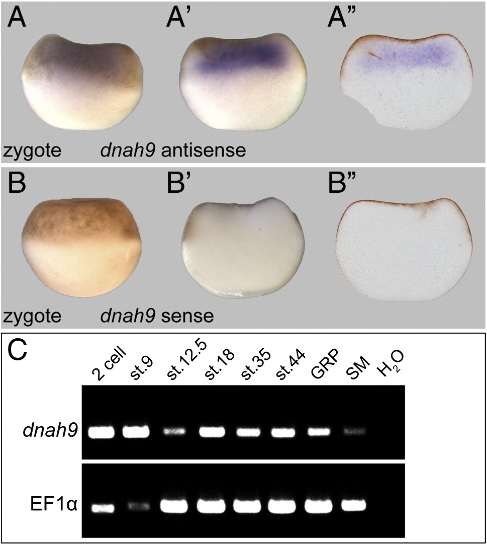
Suppl. Fig. 3. Specificity of early maternal expression patterns of dnah9. (A, B) Whole-mount in situ hybridization of zygotes using antisense (A) and sense (B) probes, shown in whole-mounts (A, B), hemi-sections (Aâ, Bâ) and 30 μm vibratome sections (Aâââ, Bâââ). (C) Semi-quantitative RT-PCR from 2-cell to tadpole stages, and of isolated GRP and SM tissue (top). Elongation factor 1 alpha (bottom) served as control. GRP, gastrocoel roof plate; SM, superficial mesoderm. Note the strong maternal expression of dnah9.
Image published in: Vick P et al. (2009)
Copyright © 2009. Image reproduced with permission of the Publisher, Elsevier B. V.
| Gene | Synonyms | Species | Stage(s) | Tissue |
|---|---|---|---|---|
| dnah9.L | dnah9-A, Left-Right Dynein, LRD, L-R Dynein | X. laevis | Throughout NF stage 1 | animal hemisphere |
Image source: Published
Permanent Image Page
Printer Friendly View
XB-IMG-154546