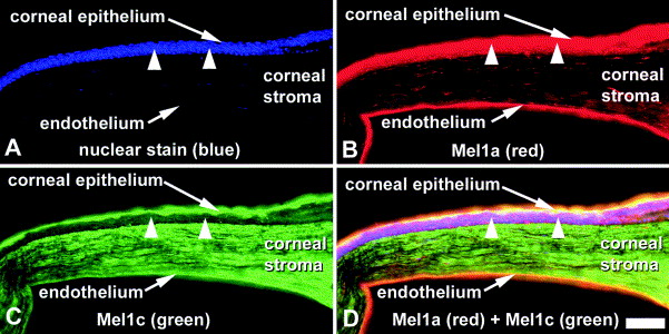
Fig. 1. Immunocytochemistry of Xenopus laevis cornea with melatonin Mel1a and Mel1c receptor antibodies. (A) Corneal section stained with blue nuclear dye (DAPI). (B) Corneal section incubated with Mel1a receptor antibody followed by incubation in secondary antibody conjugated to a red fluorescent dye. Mel1a labelling is intense in corneal epithelium (arrowheads) and endothelium, and to a lesser extent labels cells in the corneal stroma. (C) Corneal section incubated with Mel1c receptor antibody followed by incubation in secondary antibody conjugated to a green fluorescent dye. Mel1c labelling is intense in the superficial layer of corneal epithelium and in cells of the corneal stroma. The corneal endothelium and deeper layers of epithelium (arrowheads) were weakly stained. (D) Merged image of Mel1a and Mel1c labelled cornea. Yellow color indicates areas of co- localization of Mel1a and Mel1c. Scale bar=100 μm.
Image published in: Wiechmann AF and Rada JA (2003)
Copyright © 2003. Image reproduced with permission of the Publisher, Elsevier B. V.
Permanent Image Page
Printer Friendly View
XB-IMG-154998