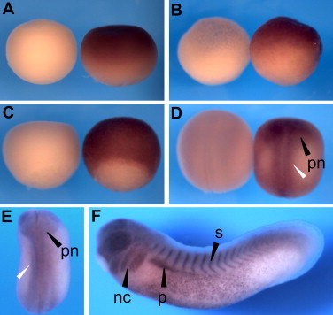
Fig. 1. In situ hybridization shows that PlexinA1 is expressed in Xenopus laevis NC cells. (AâC) PlexinA1 is expressed in the animal hemisphere of a one-cell stage embryo (A), a blastula stage 8 embryo (B) and a gastrula stage 10.5 embryo (C). Sense controls are shown on the left. (D, E) PlexinA1 expression is detected in the area of the closing neural tube (white arrow) and in premigratory NC cells at neurula stage 16 (D) and neurula stage 23 (E). (F) At tadpole stages PlexinA1 is expressed in the migrating NC cells, the eye, the pronephros and the somites. Abbreviations: nc, neural crest; pn, premigratory NC cells; p, pronephros; s, somites.
Image published in: Wagner G et al. (2010)
Copyright © 2010. Image reproduced with permission of the Publisher, Elsevier B. V.
| Gene | Synonyms | Species | Stage(s) | Tissue |
|---|---|---|---|---|
| plxna1.L | X. laevis | Throughout NF stage 1 | animal pole animal | |
| plxna1.L | X. laevis | Throughout NF stage 16 | neural plate neural fold premigratory neural crest cell cranial neural crest | |
| plxna1.L | X. laevis | Throughout NF stage 23 | cranial neural crest neural tube | |
| plxna1.L | X. laevis | Throughout NF stage 28 | neural crest cranial neural crest pharyngeal arch mandibular arch hyoid arch branchial arch eye brain somite pronephric mesenchyme pronephric kidney | |
| plxna1.L | X. laevis | Sometime during NF stage 8 to NF stage 10.5 | animal hemisphere |
Image source: Published
Permanent Image Page
Printer Friendly View
XB-IMG-156481