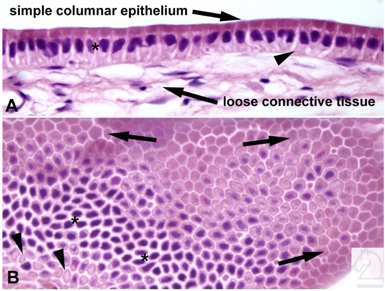
A. Simple columnar epithelium lines the lumen of the gall bladder. The columnar cells are taller than they are wide. The epithelium is observed in a longitudinal orientation. Loose connective tissue underlies the epithelium, as it does in all organs. B. Simple columnar epithelial cells of the gall bladder shown in oblique section. Image from AF Wiechmann and CR Wirsig (2003) "Color Atlas of Xenopus laevis Histology", (page 2, Chapter 1, Basic tissues: Figure 4). Copyright 2003. Kluwer Academic Publishers. Reproduced with kind permission from Springer Science & Business Media B.V.
Image published in: Color Atlas of Xenopus laevis Histology
Permanent Image Page
Printer Friendly View
XB-IMG-158225