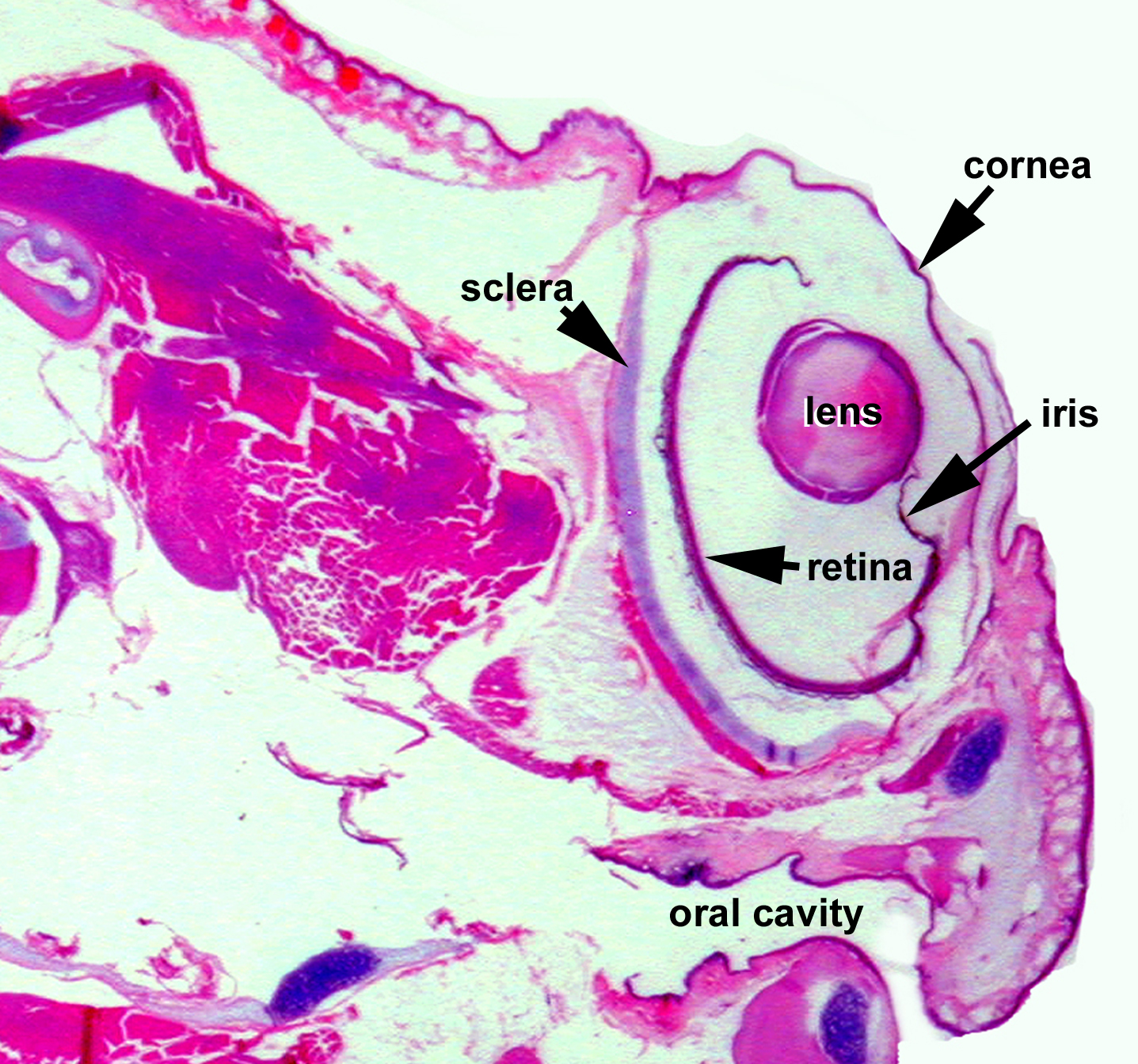
Low magnification image of the eye of a Xenopus in parasagittal plane, showing the outer surface the eye, the cornea, the position of the lens, iris and retina. The sclera contains a cartilaginous layer that adds extra support to the eye. Image from AF Wiechmann and CR Wirsig (2003) "Color Atlas of Xenopus laevis Histology", (page 122, Chapter 10: Cranial structures: Figure 61). Copyright 2003. Kluwer Academic Publishers. Reproduced with kind permission from Springer Science & Business Media B.V.
Image published in: Color Atlas of Xenopus laevis Histology
Permanent Image Page
Printer Friendly View
XB-IMG-158227