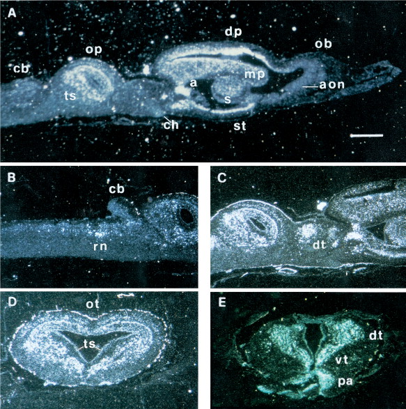
Fig. 5. Dark-field photomicrographs showing the distribution of X5-HT1A receptor mRNA in sagittal (AâC) and transverse sections (D,E) of Xenopus adult brain as revealed by in situ hybridization. In the sagittal section shown in A, the following regions appear labeled: dorsal pallium (dp), medial pallium (mp), septum (s), striatum (st), amygdala (a) in the telencephalon; optic tectum (op) and torus semicircolaris (ts) in the mesencephalon; the cerebellum (cb) as well as the olfactory bulb (ob) and the anterior olfactory nucleus (aon) are not labeled; ch, optic chiasma. (B) Sagittal section of the brain, showing the raphe nuclei (rn) of medulla oblongata labeled; the cerebellum (cb) is clearly not labeled. (C) Sagittal section of the brain showing the anterior, medial, posterior nuclei of the dorsal thalamus (dt) labeled. (D) Transverse section of mesencephalon showing the layer 6 of optic tectum (ot) and the torus semicircularis (ts) intensely labeled. (E) Transverse section of diencephalon: dorsal (dt), ventral (vt) thalamus and the preoptic area (pa) of hypothalamus are labeled. Scale bars=125 μm (in A); =150 μm (in B); =180 μm (in C); =250 μm (in D,E).
Image published in: Marracci S et al. (1997)
Copyright © 1997. Image reproduced with permission of the Publisher.
| Gene | Synonyms | Species | Stage(s) | Tissue |
|---|---|---|---|---|
| htr1a.L | 5-ht1a, 5ht1a, adrb2rl1, adrbrl1 | X. laevis | Throughout NF stage 66 | brain diencephalon midbrain dorsal pallium medial pallium telencephalon septum amygdala optic tectum telencephalon torus semicircularis striatum thalamus dorsal thalamus ventrolateral thalamic nucleus ventromedial thalamic nucleus preoptic area |
Image source: Published
Permanent Image Page
Printer Friendly View
XB-IMG-158730