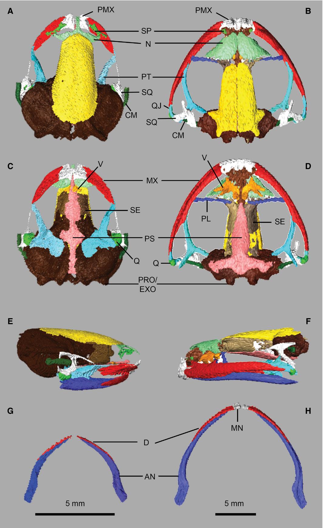
Figure 2. Skull osteology of Xenopus laevis (A,C,E,G) and Kassina maculata (B,D,F,H). Crania (upper jaw) in dorsal (A,B) and ventral (C,D) views; skull and lower jaw in lateral view (E,F); lower jaw in dorsal view (G,H). AN, angulosplenial; CM, columnella; D, dentary; MN, mentomeckelian; MX, maxilla; N, nasal; PL, palatine; PMX, premaxilla; PRO/EXO, prootic-exoccipital; PS, parasphenoid; PT, pterygoid; Q, quadrate; QJ, quadratojugal; SE, sphenethmoid; SP, septomaxilla; SQ, squamosal; V, vomer.
Image published in: Porro LB and Richards CT (2017)
Copyright © 2017. The Authors, Images redisplayed with permission of the Anatomical Society
Permanent Image Page
Printer Friendly View
XB-IMG-158973