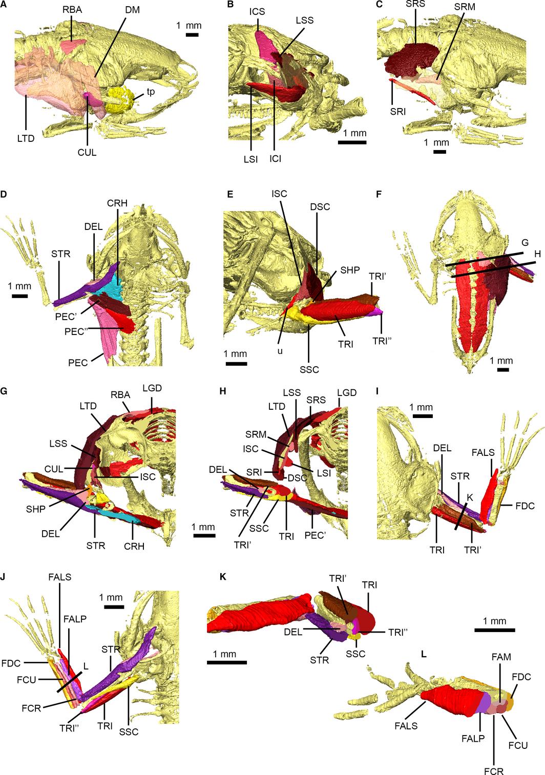
Figure 6. Pectoral and forelimb musculature of Xenopus laevis. Dorsal pectoral musculature shown in right dorsolateral view (A) with M. depressor mandibulae and M. latissimus dorsi transparent, right posterolateral view (B) with right suprascapula transparent, and right dorsolateral view (C) with suprascapula transparent. Ventral pectoral and arm musculature shown in ventral (D) and posterior (E) views. Dorsal view of specimen (F) detailing locations of transverse cross-sections through pectoral musculature (G,H). Arm and forearm musculature in dorsal (I) and ventral (J) views, and transverse cross-sections through the arm (K) and forearm (L), with sections shown in (I) and (J). Main muscles are identified using uppercase abbreviations; muscle slips and non-muscle structures are identified using lowercase abbreviations. CRH, M. coracohumeralis; CUL, M. cucullaris; DEL, M. deltoideus; DM, M. depressor mandibulae; DSC, M. dorsalis scapulae; FAM, M. flexor antebrachii medius; FALP, M. flexor antebrachii lateralis profundus; FALS, M. flexor antebrachii lateralis superficialis; FCR, M. flexor carpi radialis; FCU, M. flexor carpi ulnaris; FDC, M. flexor digitorum communis; ICI, M. intertransversarius capitis inferior; ICS, M. intertransversarius capitis superior; ISC, M. interscapularis; LGD, M. longissimus dorsi; LSI, M. levator scapulae inferior; LSS, M. levator scapulae superior; LTD, M. latissimus dorsi; PEC, M. pectoralis pars abdominalis; PEC′, M. pectoralis pars anterior sternalis; PEC″, M. pectoralis pars posterior sternalis; RBA, M. rhomboideus anterior; SHP, M. scapulo-humeralis profundus posterior; SRI, M. serratus inferior; SRM, M. serratus medius; SRS, M. serratus superior; SSC, M. subscapularis; STR, M. sternoradialis; tp, tympanic capsule; TRI, M. triceps brachii long head; TRI′, M. triceps brachii outer head; TRI″, M. triceps brachii inner head; u, unidentified pectoral girdle muscle.
Image published in: Porro LB and Richards CT (2017)
Copyright © 2017. The Authors, Images redisplayed with permission of the Anatomical Society
Permanent Image Page
Printer Friendly View
XB-IMG-158977