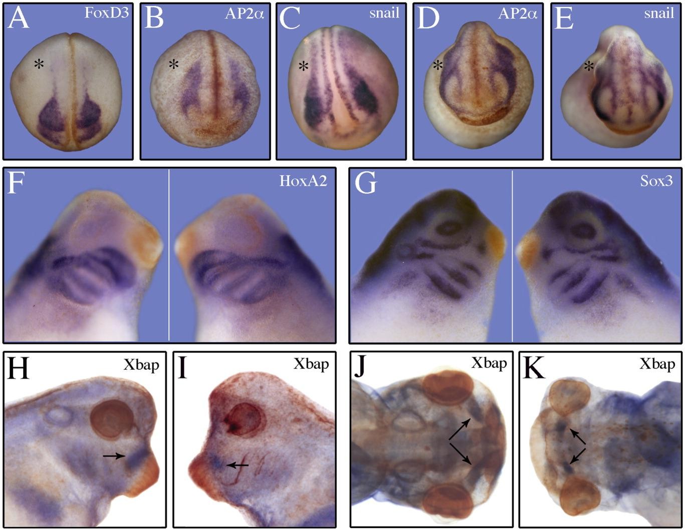
Figure 6. Effects of FoxN3 depletion on neural crest formation. Embryos were unilaterally injected with 13 ng FoxN3-MO and 500 pg GFP RNA. After sorting for right/left injections, embryos were submitted at different developmental stages to in situ whole mount hybridization. The asterisks indicate the side of injection. A: FoxD3, stage 23. B:AP2 alpha , stage 23. C:snail, stage 23. D:AP2 alpha , stage 29. E:snail, stage 29. No difference in the expression domains between injected and uninjected sides can be observed. F,G: Whole mount in situ hybridization of unilaterally injected embryos stained for HoxA2 (F) and Sox3 (G). Embryos were injected at their right sides. H-K: Whole mount in situ hybridization of bilaterally FoxN3-MO or Co-MO-injected embryos (13 ng pro blastomere). H:Xbap, Co-MO-injected, stage 38. I:Xbap, FoxN3-MO injected, stage 38. Xbap expression is visible close to the cement gland in the controls (see arrow). The FoxN3-MO-injected embryos show reduced expression of Xbap in the prospective jaw (see arrow). The ventral part of this expression domain is completely absent. J:Xbap, Co-MO-injected, stage 43, ventral view. K:Xbap, FoxN3-MO-injected, stage 43, ventral view. The Xbap expression in the controls is restricted at stage 43 within the head to Meckel's cartilage. FoxN3-MO-injected embryos show severe reduction of staining (see arrows).
Image published in: Schuff M et al. (2007)
Copyright © 2007. Image reproduced with permission of the Publisher, John Wiley & Sons.
| Gene | Synonyms | Species | Stage(s) | Tissue |
|---|---|---|---|---|
| snai1.S | LOC108702592, sna, snail, snail1, xSna, Xsnail | X. laevis | Throughout NF stage 23 | neural crest neural plate |
| tfap2a.L | ap2, AP2a, AP2alpha, tfap2a-a, tfap2a-b, tfap2alpha, XAP-2, xap2 | X. laevis | Throughout NF stage 23 | periocular region cranial neural crest |
| foxd3.L | fkh6, foxd3-a, foxd3-b, XFD-6, xfoxd3 | X. laevis | Throughout NF stage 23 | cranial neural crest neural crest eye primordium |
| snai1.S | LOC108702592, sna, snail, snail1, xSna, Xsnail | X. laevis | Throughout NF stage 23 | cranial neural crest neural plate border eye primordium |
| sox3.S | Sry, Xlsox3, xSox3, Xtsox3 | X. laevis | Sometime during NF stage 28 to NF stage 32 | otic vesicle olfactory placode lateral line placode lens brain mandibular crest hyoid crest anterior branchial crest posterior branchial crest intermediate mesoderm cranial placode |
| hoxa2.L | hox1k, hoxa2-a, hoxa2-b, hoxa2b, X-Hoxa2 | X. laevis | Sometime during NF stage 28 to NF stage 32 | hindbrain mandibular crest hyoid crest posterior branchial crest anterior branchial crest |
| tfap2a.L | ap2, AP2a, AP2alpha, tfap2a-a, tfap2a-b, tfap2alpha, XAP-2, xap2 | X. laevis | Throughout NF stage 29 and 30 | cranial neural crest periocular region head mesenchyme |
| snai1.S | LOC108702592, sna, snail, snail1, xSna, Xsnail | X. laevis | Throughout NF stage 29 and 30 | eye primordium brain cranial neural crest |
| nkx3-2.S | bagpipe homeobox homolog 1, bapx1, nkx3.2, Xbap | X. laevis | Sometime during NF stage 37 and 38 to NF stage 43 | pharyngeal arch foregut lateral plate mesoderm cartilage tissue Meckel's cartilage optic vesicle |
Image source: Published
Permanent Image Page
Printer Friendly View
XB-IMG-1682