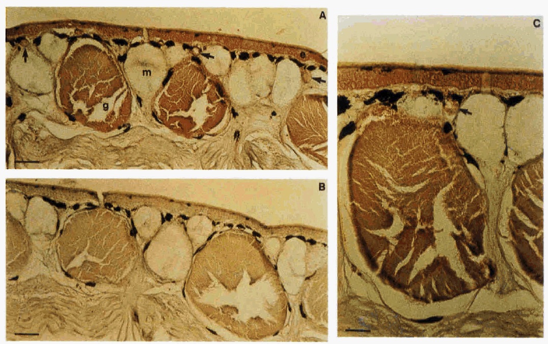
FIG. 5. Immunohistochemical analysis of X. luevis skin. A and C, positive staining (brown) of the epidermis (e), granular glands (g), and regenerating granular glands (arrows) with antiserum SKP-2. Mucous glands (m) show no immunoreactivity. B, no signals were obtained after stainiig with the preimmune serum. Sc& bars are 100 pm (A and B) and 50 pm (C).
Image published in: Hauser F et al. (1992)
Copyright © 1992. Image reproduced with permission of the Publisher.
| Gene | Synonyms | Species | Stage(s) | Tissue |
|---|---|---|---|---|
| tff3.7.S | apeg, p2, xP2 | X. laevis | Throughout NF stage 66 | skin epidermis skin gland granular gland |
Image source: Published
Permanent Image Page
Printer Friendly View
XB-IMG-172349