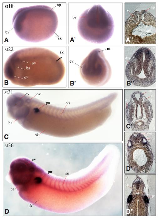
Fig. 2. Spatial analyses of lrpap1 expression. Whole-mount and sectioned in situ hybridization of wild type embryos at developmental stages 18 to 36. Earliest lrpap1 expression was detectable at NF stage 18 when neurulation starts in the neural plate and the developing brain vesicles (A,Aâ,Aââ). At NF stage 22 additional expression could be detected in the eye vesicle, the otic vesicle and the branchial arches (B,Bâ,Bââ). This expression pattern is maintained until NF stage 36 (D,Dâ,), but starting at NF stage 31 additional expression was detected in the somites and in distinct cells scattered all over the skin. Highest expression levels were observed in the proximal part of the developing pronephros (C,Câ,D). At NF stage 36 lrpap1 transcripts are spread through the entire pronephric epithelium and the developing otic vesicles (D,Dâ,Dââ). Abbreviations: (bv) brain vesicle, (ba) branchial arches, (dev) distal eye vesicle, (dnt) dorsal neural tube, (ep) epithelium, (ev) eye vesicle, (np) neural plate, (nt) neural tube, (ov) otic vesicle, (pn) pronephros, (sk) skin, (so) somites. A, B, C and D show lateral views of the embryos, Aâ and Bâ show frontal views.
Image published in: Neuhaus H et al. (2018)
Copyright © 2018. Image reproduced with permission of the Publisher, University of the Basque Country Press.
| Gene | Synonyms | Species | Stage(s) | Tissue |
|---|---|---|---|---|
| lrpap1.L | X. laevis | Sometime during NF stage 18 to NF stage 28 | neural plate pre-chordal neural plate chordal neural plate | |
| lrpap1.L | X. laevis | Throughout NF stage 22 | optic vesicle cranial neural crest neural tube posterior neural tube anterior neural tube otic placode | |
| lrpap1.L | X. laevis | Throughout NF stage 31 | eye pronephric nephron pronephric tubule proximal tubule neural tube pharyngeal arch somite | |
| lrpap1.L | X. laevis | Throughout NF stage 35 and 36 | eye pronephric kidney pronephric nephron pronephric tubule proximal tubule somite brain skin pharyngeal arch |
Image source: Published
Permanent Image Page
Printer Friendly View
XB-IMG-172981