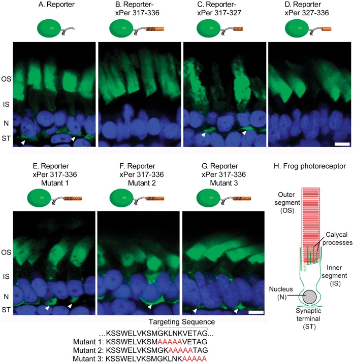
Figure 1. The peripherin targeting signal is contained within a ten amino acid residue stretch. Panels show confocal images of transgenic frog retinas expressing the reporter construct YFP-xRhoCTδ5 (green) fused to the fragments of the peripherin C-terminus illustrated in cartoons above the corresponding panels. Partial mislocalization of several constructs from rod outer segments is marked by white arrowheads. (A) The YFP-xRhoCTδ5 reporter. (B) The reporter fused to xPer 317â336. (C) The reporter fused to xPer 317â327. (D) The reporter fused to xPer 327â336. (EâG) The reporter fused to xPer 317â336 sequences containing polyalanine amino acid substitutions indicated below the panels. Abbreviations are: OS â outer segment, IS â inner segment, N â nuclei, ST â synaptic termini. The nuclei (blue) are stained with Hoechst; scale bar: 5 µm. (H) A schematic of a frog rod photoreceptor illustrating its principle compartments.
Image published in: Salinas RY et al. (2013)
Image reproduced on Xenbase with permission of the publisher and the copyright holder. Creative Commons Attribution license
Permanent Image Page
Printer Friendly View
XB-IMG-173568