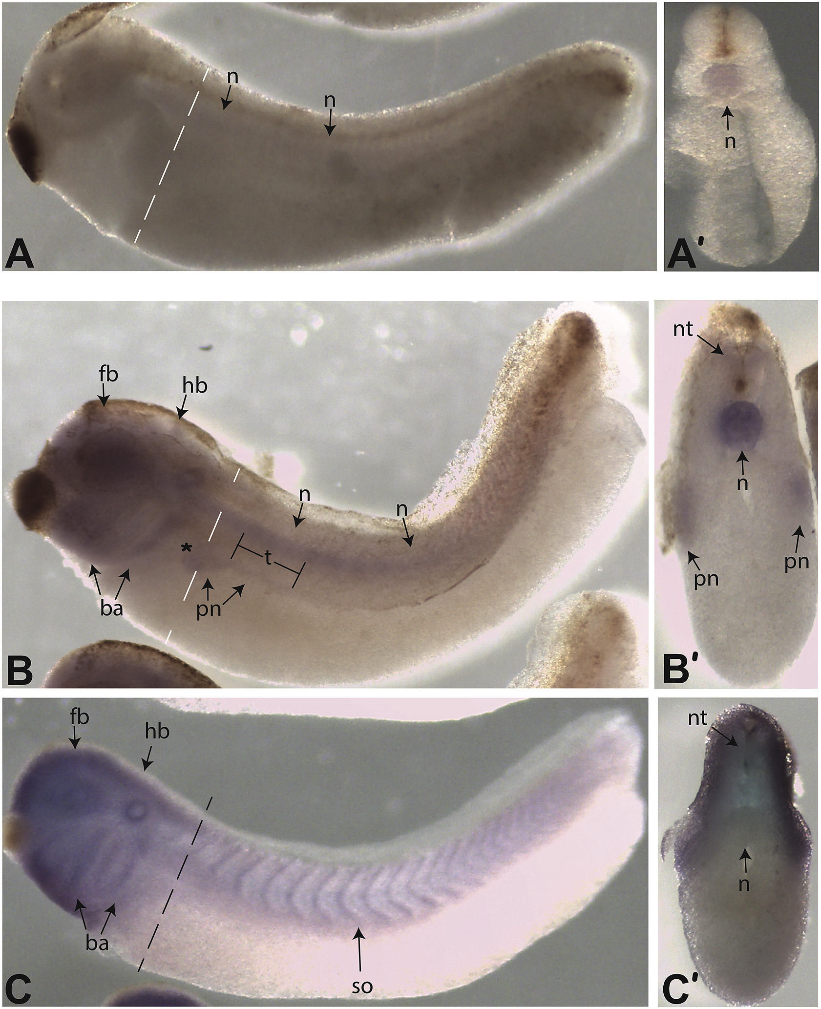
Fig. 4. The expression of gtpbp10 in late stages of development shown by whole-mount in situ hybridization (WISH). gtpbp10 antisense riboprobe was used for in situ hybridization on A) Early tail bud stage embryo (stage 28) and B) Late tail-bud stage (stage 36). Aâ² and Bâ² are transverse sections of the embryos shown in A and B at the level of the dotted white lines. Structures labeled as follows: notochord (n), neural tube (nt), forebrain (fb), hindbrain (hb), pronephros (pn), branchial arches (ba). * denotes the glomerulus and proximal tubules, while âtâ indicates the distal tubules. C) gtpbp10 sense probe shows background staining in the anterior branchial arches, otic vesicle, eye, and fore-, mid-, and hindbrain. Staining was also observed in somitic tissue (so) along the anterior-posterior axis.
Image published in: Jerry R et al. (2019)
Copyright © 2019. Image reproduced with permission of the Publisher, Elsevier B. V.
| Gene | Synonyms | Species | Stage(s) | Tissue |
|---|---|---|---|---|
| gtpbp10.L | X. laevis | Throughout NF stage 28 | notochord | |
| gtpbp10.L | X. laevis | Throughout NF stage 35 and 36 | notochord central nervous system spinal cord pronephric kidney |
Image source: Published
Permanent Image Page
Printer Friendly View
XB-IMG-175272