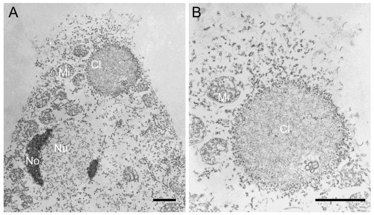
Figure 2. (A,B) Transmission electron microscopy of permeabilized Dictyostelium cells showing spongy GFP-NE81ΔNLSΔCLIM clusters (Cl) studded by particles representing ribosomes. The nucleus (Nu), nucleoli (No), and mitochondria (Mi) are labeled. (B) is an enlarged view of (A). Scale bars = 1 µm.
Image published in: Grafe M et al. (2019)
© 2019 by the authors. Creative Commons Attribution license
Permanent Image Page
Printer Friendly View
XB-IMG-175330