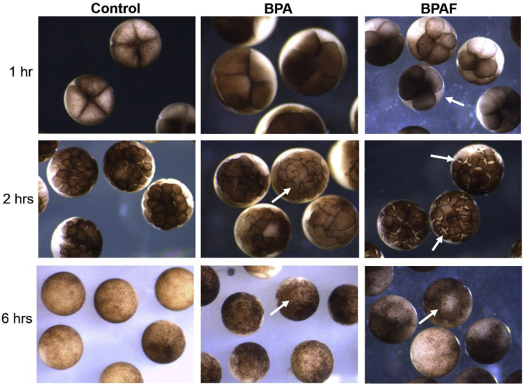
Fig. 1. Early cleavage division defects caused by BPA and BPAF. Control embryos (left column), compared to embryos exposed to BPA (50 μM; middle column) or BPAF (3 μM; right column) at 1, 2 and 6 hours of exposure. Irregular, asymmetrical mitotic division, slowed cytokinesis, and cellular dissociation were observed in the majority of BPA- and BPAF-treated embryos (arrows).
Image published in: Arancio AL et al. (2019)
© 2019 The Author(s). Creative Commons Attribution license
Permanent Image Page
Printer Friendly View
XB-IMG-176246