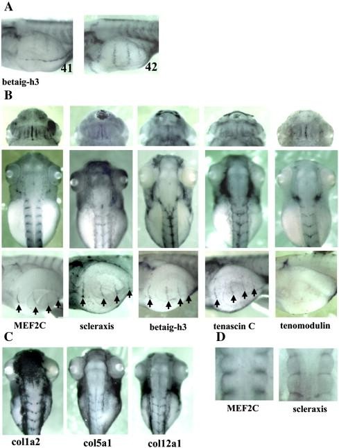
Fig. 5. Comparison of the localization of XMEF2C, Xscleraxis, betaig-h3, tenascin C and tenomodulin mRNA by whole-mount in situ hybridization. (A) Betaig-h3 mRNA begins to be expressed in connective tissue associated with hypaxial muscle at stage 41. (B) XMEF2C, Xscleraxis, betaig-h3, tenascin C and tenomodulin mRNA are largely colocalized at stage 45 in head region (ventral view, top) intersomitic space as indicated by staining between the convex myotome (dorsal view, middle). Connective tissue associated with hypaxial muscles expresses XMEF2C, Xscleraxis, betaig-h3 and tenascin C, but we cannot detect tenomodulin mRNA (lateral view, bottom). (C) Col1a2, col5a1 and col12a1 mRNA is also expressed in intersomitic space at stage 45 (D) Comparison of MEF2C and scleraxis mRNA accumulation 3 days after feeding. MEF2C mRNA is always expressed at this stage while scleraxis mRNA appears stronger. Arrows indicate connective tissue associated with hypaxial muscles expression. Numbers indicate developmental stages.
Image published in: della Gaspera B et al. (2009)
Copyright © 2009. Image reproduced with permission of the Publisher, Elsevier B. V.
| Gene | Synonyms | Species | Stage(s) | Tissue |
|---|---|---|---|---|
| tgfbi.L | betaig-h3, bigh3, cdb1, cdg2, cdgg1, csd, csd1, csd2, csd3, ebmd, keratoepithelin, lcd1, LOC108712407, xtgfbi | X. laevis | Sometime during NF stage 41 to NF stage 42 | tendon somite hypaxial muscle connective tissue |
| scx.L | scleraxis, xscleraxis | X. laevis | Throughout NF stage 45 | muscle head region tendon somite hypaxial muscle connective tissue |
| tnc.L | LOC108700035, tenascin, tenascin C, tenascin-C | X. laevis | Throughout NF stage 45 | hypaxial muscle connective tissue tendon |
| mef2c.L | XMEF2C | X. laevis | Throughout NF stage 45 | head region muscle tendon myotome somite hypaxial muscle connective tissue |
| tnmd.S | X. laevis | Throughout NF stage 45 | tendon somite connective tissue | |
| col1a2.L | MGC52958, procollagen, type I, alpha 2 | X. laevis | Throughout NF stage 45 | tendon somite connective tissue |
| col5a1.L | X. laevis | Throughout NF stage 45 | muscle tendon somite connective tissue | |
| col12a1.L | X. laevis | Throughout NF stage 45 | muscle tendon somite hypaxial muscle connective tissue |
Image source: Published
Permanent Image Page
Printer Friendly View
XB-IMG-30705