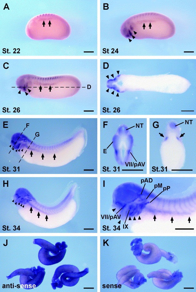
Fig. 3. Spatial expression pattern of X. laevis GDNF in developing embryos. Whole-mount in situ hybridization analysis showed the localization of X. laevis GDNF transcripts. Lateral view at stage 22 (A), stage 24 (B), stage 26 (C), stage 31 (E), and stage 34 (H and I). Dorsal is up, and anterior is left. (D) The longitudinal section at stage 26. Anterior is left. (F and G) Transverse sections at stage 31 in the eye (F) and the pronephros (G) levels. Dorsal is up. (J and I) Digestive tracts dissected from stage 45 embryos hybridized with anti-sense (J) and sense (K) probe. Arrows and arrowheads indicate pronephros and pharyngeal arches, respectively. The dotted line in (C) illustrates the plane of section in (D). The dotted lines through the eye and the pronephros in (E) correspond to planes of section in (F) and (G), respectively. Abbreviations: E, eye; IX, glossopharyngeal epibranchial placode; NT, neural tube; pAD, anterodorsal lateral line placode; pM, middle lateral line placode; pP, posterior lateral line placode; VII/pAV, a composite structure of the facial epibranchial placode and the anteroventral lateral line placode. Scale bar is 500Â micrometers.
Image published in: Kyuno J and Jones EA (2007)
Copyright © 2007. Image reproduced with permission of the Publisher, Elsevier B. V.
| Gene | Synonyms | Species | Stage(s) | Tissue |
|---|---|---|---|---|
| gdnf.L | gdnf-a, gdnf-b, hfb1-gdnf | X. laevis | Throughout NF stage 22 | pronephric mesenchyme |
| gdnf.L | gdnf-a, gdnf-b, hfb1-gdnf | X. laevis | Sometime during NF stage 24 to NF stage 33 and 34 | pronephric kidney pharyngeal arch |
| gdnf.L | gdnf-a, gdnf-b, hfb1-gdnf | X. laevis | Throughout NF stage 31 | eye neural tube epibranchial placode anterior ventral lateral line placode |
| gdnf.L | gdnf-a, gdnf-b, hfb1-gdnf | X. laevis | Throughout NF stage 33 and 34 | pronephric kidney pharyngeal arch anterior dorsal lateral line placode middle lateral line posterior epibranchial placode ventral |
Image source: Published
Permanent Image Page
Printer Friendly View
XB-IMG-42721