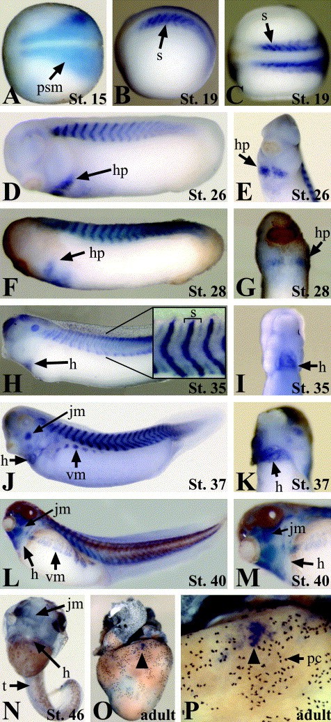
Fig. 2. Xtn3 is developmentally expressed throughout the somites, heart, and jaw myoblasts from early neurula to late tailbud stages. (AâP) In situ hybridization showing expression of the Xtn3 at the indicated stages. (AâC) Neurula stage embryos. Anterior is to the left. (A and C) Dorsal views. (B) Lateral view. (DâM) Tailbud stage embryos. (E, G, I, K, and M) Ventral anterior views. Anterior is up. (H) Inset shows higher magnification of somite region. Bracket indicates one individual somite. (N) Tadpole stage embryo. Note the lack of staining in trunk and heart. Some light staining remains in facial muscles. (O and P) Adult heart lacking global Xtn3 expression. (P) A magnification of (O). Black arrowhead indicates isolated group of Xtn3-expressing cells. h, heart; hp, heart primordia; jm, jaw myoblasts; psm, presomitic mesoderm; pc, pigment cells; vm, ventral myoblasts; s, somites; t, trunk.
Image published in: Brown DD et al. (2006)
Copyright © 2006. Image reproduced with permission of the Publisher, Elsevier B. V.
| Gene | Synonyms | Species | Stage(s) | Tissue |
|---|---|---|---|---|
| tn3.L | Xtn3 | X. laevis | Throughout adult frog stage | myocardium |
| tn3.L | Xtn3 | X. laevis | Throughout NF stage 15 | paraxial mesoderm |
| tn3.L | Xtn3 | X. laevis | Throughout NF stage 19 | somite presomitic mesoderm |
| tn3.L | Xtn3 | X. laevis | Throughout NF stage 26 | heart primordium somite presomitic mesoderm |
| tn3.L | Xtn3 | X. laevis | Throughout NF stage 28 | somite cardiac mesoderm heart |
| tn3.L | Xtn3 | X. laevis | Throughout NF stage 35 and 36 | somite otic vesicle heart myocardium cardiac mesoderm mesoderm intersomitic region mesenchyme |
| tn3.L | Xtn3 | X. laevis | Throughout NF stage 37 and 38 | jaw muscle somite mesenchyme myoblast abdominal myoblast heart cardiac mesoderm |
| tn3.L | Xtn3 | X. laevis | Throughout NF stage 40 | myocardium somite heart myoblast abdominal myoblast musculature of face jaw muscle |
| tn3.L | Xtn3 | X. laevis | Throughout NF stage 46 | musculature of face |
Image source: Published
Permanent Image Page
Printer Friendly View
XB-IMG-42738