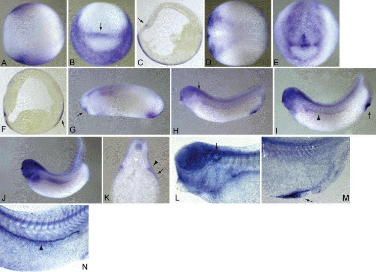
Fig. 2. Expression of Xsl-1 RNA. All embryos are pictured with the anterior to the left unless otherwise noted (B,E,K). Stages 12 (AâC) and 17 (DâE) are shown from dorsal views (A,D), frontal views (B,E), where dorsal is to the top of the frame, and as saggital sections (C,F). Stages 24 (G), 28 (H), 33 (I,L,M), and 37 (J,N) are shown as lateral views. Panel K is a transverse section of a stage 37 embryo at approximately the level of fourth trunk somite. High magnification views of a cleared stage 33 embryo are pictured in L (anterior) and M (posterior). A high magnification view of expression in the pronephros, somites, and neural tube of a cleared stage 37 embryo is seen in N. Further description of the arrows and arrowheads can be found in Section 1.
Image published in: Martin BL and Harland RM (2004)
Copyright © 2004. Image reproduced with permission of the Publisher, Elsevier B. V.
| Gene | Synonyms | Species | Stage(s) | Tissue |
|---|---|---|---|---|
| kitlg | steel, Xkl-1, Xsl, Xsl-1, Xsl-2 | Xenopus | Throughout NF stage 12 | epidermis outer layer ectoderm neuroectoderm inner layer |
| kitlg | steel, Xkl-1, Xsl, Xsl-1, Xsl-2 | Xenopus | Sometime during NF stage 12 to NF stage 37 and 38 | epidermis |
| kitlg | steel, Xkl-1, Xsl, Xsl-1, Xsl-2 | Xenopus | Sometime during NF stage 17 to tadpole stage | epidermis cement gland primordium proctodeum |
| kitlg | steel, Xkl-1, Xsl, Xsl-1, Xsl-2 | Xenopus | Sometime during NF stage 28 to NF stage 66 | |
| kitlg | steel, Xkl-1, Xsl, Xsl-1, Xsl-2 | Xenopus | Throughout NF stage 37 and 38 | pronephric duct dorsal neural tube |
| kitlg | steel, Xkl-1, Xsl, Xsl-1, Xsl-2 | Xenopus | Throughout NF stage 45 to NF stage 66 | pronephric kidney neural tube otic placode pharyngeal arch somite |
Image source: Published
Permanent Image Page
Printer Friendly View
XB-IMG-42870