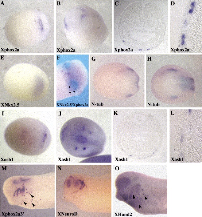
Fig. 3. Ventral and lateral domains of Xphox2a and Xash1 expression. Patterns of gene expression were characterized as indicated by whole-mount in situ hybridization at neural plate/fold (AâE, GâL), neurula (F) and early tailbud stages (MâO). The ventral side of the embryo is shown in (A,EâG,I). In (B,H,J), both dorsal and ventral domains of XPhox2a, N-tubulin and Xash1 expression are visible in a neural plate/fold stage embryo. In (F), Xnkx2.5 staining is blue and Xphox2a positive cells are magenta. A transverse section through the neural plate of embryos probed for Xphox2a(C,D) or Xash1(K,L) is shown at low (C,K) and high (D,L) magnifications.
Image published in: Talikka M et al. (2004)
Copyright © 2004. Image reproduced with permission of the Publisher, Elsevier B. V.
| Gene | Synonyms | Species | Stage(s) | Tissue |
|---|---|---|---|---|
| nkx2-5.L | AR2, csx, nkx-2.5, Nkx-2.5, nkx2.5, nkx2-5-a, nkx2-5-b, tinman, XNkx-2.5, XNkx2-5 | X. laevis | Throughout NF stage 14 | cardiac progenitor cell anterior ventral mesoderm |
| tubb2b.S | NBT, neural beta-tubulin, NST, N-Tub, n-tubulin, ntubulin, Xn-tubulin | X. laevis | Throughout NF stage 14 | neural plate lateral chordal neural plate posterior |
| phox2a.S | LOC108710089, phox2, xphox2a | X. laevis | Throughout NF stage 14 to NF stage 21 | anterior ventral mesoderm cardiac progenitor cell |
| ascl1.L | ash1, Mash1, Xash1 | X. laevis | Throughout NF stage 14 to NF stage 21 | ventral anterior mesoderm cardiac progenitor cell |
| hand2.L | dhand, dhand2, hand2-a, hand2-b, xhand2 | X. laevis | Throughout NF stage 22 to NF stage 28 | smooth muscle pericyte |
| neurod1.L | neurod, neuroD, neurod1-a, neurod1-b, XNeuroD | X. laevis | Throughout NF stage 28 | neuron lens placode eye primordium lateral line placode olfactory placode trigeminal placode |
Image source: Published
Permanent Image Page
Printer Friendly View
XB-IMG-43488