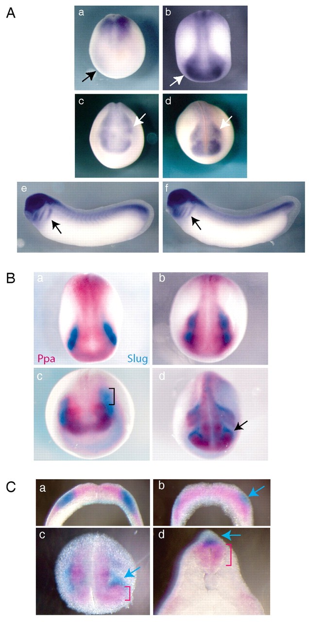
Fig. 4. Ppa is dynamically expressed in neural crest-forming regions. (A) In situ hybridization showing the developmental expression of Ppa at stage 13 (a) stage 16 (b) stage 19 (c), stage 22 (d) stage 24 (e) and stage 28 (f). Strong expression is seen in the neural crest region of late neural plate stage embryos (b,c, arrows) and in the migrating neural crest at tailbud stages (d-f, arrows). By contrast, no Ppa expression is seen in neural crest precursor-forming regions at stage 13 (a, arrow). (B) Double in situ hybridizations comparing the expression patterns of Ppa (pink) and Slug (light blue). At stages 15 (a) and 18 (b), Ppa and Slug expression is entirely overlapping. However, at stages 20 (c) and 22 (d), Ppa and Slug expression patterns are only partially coincident. Bracket in c indicates region of Slug-expressing cells that do not expess Ppa. Arrow in d indicates migratory neural crest cells that express Slug but not Ppa. (C) Vibratome sections of double in situ hybridizations demonstrate the complete colocalization of Ppa and Slug expression in the rostral region of a stage 15 embryo (a). A more caudal section from the same embryo as in a, showing that Ppa is not expressed in the newly induced, Slug-expressing neural crest precursors (b, arrow). Sections through cranial (c) and spinal cord (d) regions of a stage 24 embryo. Slug is expressed in the earliest migrating neural crest (c,d, arrow) but Ppa is excluded from these cells (c,d, bracket).
Image published in: Vernon AE and LaBonne C (2006)
Copyright © 2006. Image reproduced with permission of the Publisher and the copyright holder. This is an Open Access article distributed under the terms of the Creative Commons Attribution License.
| Gene | Synonyms | Species | Stage(s) | Tissue |
|---|---|---|---|---|
| snai2 | slug, snai2-a, snai2-b, Snail2, xSlu, xslug, XSnail2 | Xenopus | Sometime during NF stage 15 to NF stage 18 | neural plate lateral |
| fbxl14 | fbl13, Ppa | Xenopus | Sometime during NF stage 16 to NF stage 19 | neural plate lateral |
| snai2 | slug, snai2-a, snai2-b, Snail2, xSlu, xslug, XSnail2 | Xenopus | Sometime during NF stage 20 to NF stage 24 | neural crest |
| fbxl14 | fbl13, Ppa | Xenopus | Sometime during NF stage 20 to NF stage 28 | neural crest |
Image source: Published
Permanent Image Page
Printer Friendly View
XB-IMG-45766