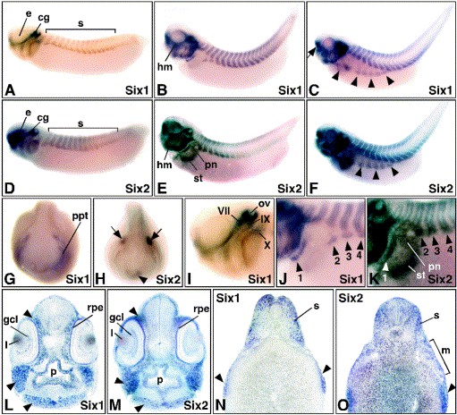
Fig. 2. Analysis of Six2 subfamily gene expression during Xenopus development by whole-mount in situ hybridization. Lateral views are shown in (AâF,IâK) with anterior to the left. Frontal views (G,H) and the sections (LâO) are oriented with dorsal to the top. (AâC) Six1 expression in stage 30 (A), stage 35 (B) and stage 39 (C) embryos. The arrow in panel (C) indicates the olfactory bulbs and the arrowheads highlight the migrating ventral muscle anlagen. (DâF) Six2 expression in stage 30 (D), stage 35 (E) and stage 39 (F) embryos. The arrowheads in panel (F) indicate ventral muscle anlagen. (G,H) Frontal views of stage 20 embryos showing Six1 (G) and Six2 (H) gene expression. The olfactory (arrowhead) and otic placodes (arrows) are indicated in panel H. (I) Close-up view of a stage 30 embryo head illustrating Six1 expression associated with facial (VII), glossopharyngeal (IX) and vagus (X) ganglia. (J,K) Close-up views of stage 35 embryos demonstrating Six1 (J) and Six2 (K) expression in the 1stâ4th ventral abdominal muscle anlagen (arrowheads). (L,M) Transverse sections through the head of stage 39 embryos illustrating Six1 (L) and Six2 (M) expression. Arrowheads highlight expression in the head mesenchyme. (N) Transverse section through the midtrunk of a stage 39 embryo. Arrowheads indicate Six1 expression in migrating abdominal muscle anlagen. (O) Transverse section at the level of the pronephric kidney through a stage 39 embryo. The abdominal muscle anlagen (arrowhead), and the mesenchyme (m) adjacent to the developing stomach and pronephric kidney epithelia are indicated. Abbreviations: cg, cranial ganglia; e, eye; gcl, ganglion cell layer; hm, head mesenchyme; l, lens; p, pharynx; pn, pronephros; ppt, primitive placodal thickening; rpe, retinal pigment epithelium; s, somites; st, stomach.
Image published in: Ghanbari H et al. (2001)
Copyright © 2001. Image reproduced with permission of the Publisher, Elsevier B. V.
| Gene | Synonyms | Species | Stage(s) | Tissue |
|---|---|---|---|---|
| six1.L | XSix1 | X. laevis | Throughout NF stage 20 | olfactory placode |
| six2.L | X. laevis | Throughout NF stage 20 | otic placode olfactory placode | |
| six1.L | XSix1 | X. laevis | Throughout NF stage 28 | somite cranial ganglion olfactory placode |
| six1.L | XSix1 | X. laevis | Throughout NF stage 29 and 30 | otic vesicle olfactory bulb facial epibranchial placode glossopharyngeal epibranchial placode vagal epibranchial placode cranial placode |
| six2.L | X. laevis | Throughout NF stage 29 and 30 | eye somite cranial ganglion intermediate mesoderm lateral plate mesoderm olfactory placode | |
| six1.L | XSix1 | X. laevis | Throughout NF stage 35 and 36 | cranial ganglion head mesenchyme somite hypaxial muscle pharyngeal region abdominal myoblast |
| six2.L | X. laevis | Throughout NF stage 35 and 36 | pronephric kidney stomach head mesenchyme otic vesicle head mesenchyme eye retina retinal ganglion cell layer pharyngeal mesenchyme olfactory placode | |
| six1.L | XSix1 | X. laevis | Throughout NF stage 39 | lens somite olfactory bulb hypaxial muscle abdominal myoblast |
| six2.L | X. laevis | Throughout NF stage 39 | head otic vesicle stomach eye pronephric kidney cranial ganglion somite olfactory bulb head mesenchyme hypaxial muscle foregut pharyngeal region |
Image source: Published
Permanent Image Page
Printer Friendly View
XB-IMG-46062