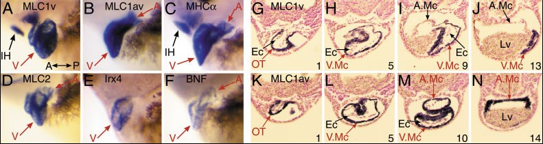
Fig. 2. The ventricular chamber-restricted embryonic expression of Xenopus MLC1v compared with other cardiac markers. High-magnification, left lateral views of the hearts of tadpoles (A, anterior to the left) that had been rendered transparent using benzyl alcohol/benzyl benzoate treatment, after whole-mount in situ hybridization. A: Cardiac MLC1v mRNA expression at stage 38. B: MLC1av expression at stage 39. C: MHC expression at stage 39. D: MLC2 expression at stage 38. E: Irx4 expression at stage 38. F: B-Type natriuretic peptide precursor (BNF) expression at stage 38. Cardiac domains of gene expression are indicated by red arrows and other domains by black arrows. The apparent differences in outflow tract gene expression of the four sarcomeric muscle proteins shown are due to the changing rostral to caudal position of the proximal outflow tract between stages 38 to 39. Only Irx4 mRNA is absent from the proximal outflow tract. IH, interhyoid facial muscle; V, ventricle; A, atria. G: Transverse sections through Xenopus stage 35 tadpole hearts after whole-mount in situ hybridization for MLC1v and MLC1av mRNA. Four representative sections (10 m) marking progressively posterior cardiac slices are shown for MLC1v (G) and MLC1av (K), with the section number indicated at the bottom-right of each panel. Ec, endocardium; OT, outflow tract; V.Mc, ventricular myocardium; A.Mc, atrial myocardium; LV, liver; MLC, myosin light chain.
Image published in: Smith SJ et al. (2005)
Copyright © 2005. Image reproduced with permission of the Publisher, John Wiley & Sons.
| Gene | Synonyms | Species | Stage(s) | Tissue |
|---|---|---|---|---|
| irx4.L | iroquois-4, irx4-a, irx4-b, irxa3, Xiro4 | X. laevis | Throughout NF stage 37 and 38 | cardiac ventricle |
| myl3.S | MLC1v, myl3-a, myl3-b, xMLC1v | X. laevis | Throughout NF stage 37 and 38 | heart cardiac ventricle jaw muscle |
| myl4.L | MLC1av | X. laevis | Throughout NF stage 37 and 38 | left atrium right atrium cardiac ventricle |
| myl7.L | mlc2, myl2a, mylc2a | X. laevis | Throughout NF stage 37 and 38 | cardiac ventricle left atrium right atrium |
| nppb.L | bnf, bnp | X. laevis | Throughout NF stage 37 and 38 | heart left atrium right atrium cardiac ventricle |
| myh6.S | aMHC, ck231, MHC, MHCa, MHC-alpha, MHCalpha, muz, muzak, XCMHC | X. laevis | Throughout NF stage 39 | jaw muscle left atrium right atrium cardiac ventricle |
Image source: Published
Permanent Image Page
Printer Friendly View
XB-IMG-47124