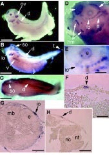
Fig. 4. Expression of xPAK during lateral line development. (A) Stage 35/36, (B-H) stage 37/38. (A-E) Whole-mount in situ hybridization. (F,G and H) Transverse sections of stage 37/38 whole mount embryos sectioned after hybridization with xPAK1. ov, otic vesicle; o, olefactory placode; nc, notochord; e, eye; a, d, io, so, hm, respectively the aortic, dorsal, infraor- bital, supraorbital and hyomandibular lateral lines; t, tail. Bar in A to C,1 mm; in D, 0.3 mm; in E and F, 0.16 mm; and in G and H, 0.3 mm.
Image published in: Islam N et al. (2000)
Copyright © 2000. Image reproduced with permission of the Publisher.
| Gene | Synonyms | Species | Stage(s) | Tissue |
|---|---|---|---|---|
| pak1.S | MGC68680, p21-activated kinase, pak, pak-1, pakalpha, X-PAK1, XPak1 | X. laevis | Throughout NF stage 35 and 36 | otic vesicle lens olfactory region dorsal lateral line lateral line placode pharyngeal arch ventral lateral line tail bud |
| pak1.S | MGC68680, p21-activated kinase, pak, pak-1, pakalpha, X-PAK1, XPak1 | X. laevis | Throughout NF stage 37 and 38 | otic vesicle lateral line system lateral line lateral line placode olfactory bulb ventral lateral line neuromast mantle cell dorsal lateral line infraorbital lateral line infraorbital lateral line primordium middle lateral line occipital lateral line supraorbital lateral line trunk lateral line eye tail bud hyomandibular lateral line pharyngeal arch |
Image source: Published
Permanent Image Page
Printer Friendly View
XB-IMG-49674