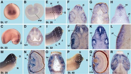
Figure 3. Horizontal and transversal section analyses of the IRS-1 expression pattern. anp, anterior neural plate; ba, branchial arches; cmz, ciliary marginal zone; di, diencephalon; g, gill; le, lens epithelium; lf, lens fibers; lpl: lens placode; opv, optic vesicle; ov, otic vesicle; pt, proximal tubule of pronephros; sc, spinal cord.Download figure to PowerPoint
Image published in: Bugner V et al. (2011)
Copyright © 2011. Image reproduced with permission of the Publisher, John Wiley & Sons.
| Gene | Synonyms | Species | Stage(s) | Tissue |
|---|---|---|---|---|
| irs1.L | irs-1, LOC108718134 | X. laevis | Throughout NF stage 17 | pre-chordal neural plate optic field |
| irs1.L | irs-1, LOC108718134 | X. laevis | Throughout NF stage 23 | eye diencephalon optic vesicle |
| irs1.L | irs-1, LOC108718134 | X. laevis | Throughout NF stage 28 | diencephalon lens placode branchial arch spinal cord |
| irs1.L | irs-1, LOC108718134 | X. laevis | Throughout NF stage 32 | lens lens placode diencephalon otic vesicle early proximal tubule |
| irs1.L | irs-1, LOC108718134 | X. laevis | Throughout NF stage 35 and 36 | lens lens epithelium lens fiber cell mass |
| irs1.L | irs-1, LOC108718134 | X. laevis | Throughout NF stage 40 | cornea lens lens epithelium lens transitional zone ciliary marginal zone external gill |
Image source: Published
Permanent Image Page
Printer Friendly View
XB-IMG-74767