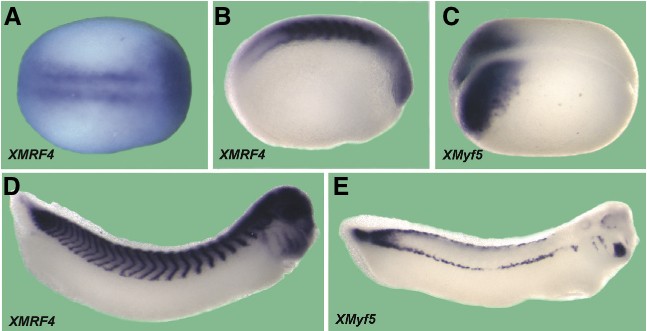
Fig. 2. Whole mount in situ hybridization of X. laevis embryos with the full-length XMRF4 probe or XMyf5 probe. All are oriented anterior to the right. (A) Stage 14- 15 early neurula, dorsal view. XMRF4 expression is seen in presomitic mesoderm prior to segmentation, as well as in the anterior. (B) Stage 20 neurula, lateral view. XMRF4 staining is most intense in the somites but also evident in the eye primordia. (C) Stage 16 neurula, dorsolateral view. XMyf5 expression is confined to the posterior mesoderm. (D) Stage 31-32 tailbud embryo, lateral view. In addition to the myotomal staining, XMRF4 expression is evident in the eyes, brain, branchial arches, otic vesicles, and head meso- derm. (E) Stage 31-32 tailbud embryo, lateral view. XMyf5 expression is seen in tailbud mesoderm, dorsal and ventral myotomal cells, and in primordia of some cranial muscles.
Image published in: Hinterberger TJ (2010)
Copyright © 2010. Image reproduced with permission of the Publisher, University of the Basque Country Press.
| Gene | Synonyms | Species | Stage(s) | Tissue |
|---|---|---|---|---|
| myf6.L | herculin, mrf4, myf6-a, myf6-b, Xmrf4, XMRF4-a, XMRF4a | X. laevis | Throughout NF stage 14 | dorsal presomitic mesoderm pre-chordal neural plate optic field |
| myf5.L | myf-5, Xmyf-5, Xmyf5 | X. laevis | Throughout NF stage 16 | mesoderm posterior |
| myf6.L | herculin, mrf4, myf6-a, myf6-b, Xmrf4, XMRF4-a, XMRF4a | X. laevis | Throughout NF stage 20 | somite presomitic mesoderm |
| myf6.L | herculin, mrf4, myf6-a, myf6-b, Xmrf4, XMRF4-a, XMRF4a | X. laevis | Throughout NF stage 31 to NF stage 32 | somite otic vesicle eye brain branchial arch head mesoderm |
| myf5.L | myf-5, Xmyf-5, Xmyf5 | X. laevis | Throughout NF stage 31 to NF stage 32 | myotome tail region head region presomitic mesoderm myoblast |
Image source: Published
Permanent Image Page
Printer Friendly View
XB-IMG-75428