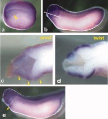
Figure 1. Determination of the expression pattern of ARVCF by whole-mount in situ hybridization. X. laevis embryos at different developmental stages were examined using an antisense probe directed against ARVCF. a: Lateral view of stage-18 embryo with dorsal side at the top, anterior to the left. The arrowhead shows enriched transcripts in the neural plate and bordering neural crest. b: Lateral view of a stage-26 tailbud with the anterior towards the left. The line shows the position of the section shown in panel c. c: Horizontal section through the head region of a stage-26 tailbud showing the presence of ARVCF transcripts in the cranial neural crest cells (yellow arrowhead). d: A similar section of a stage-26 embryo stained with the neural crest marker Twist as a reference. e: Stage-31 early tadpole showing enriched ARVCF expression in the head region and the heart (yellow arrowhead).Download figure to PowerPoint
Image published in: Tran HT et al. (2011)
Copyright © 2011. Image reproduced with permission of the Publisher, John Wiley & Sons.
| Gene | Synonyms | Species | Stage(s) | Tissue |
|---|---|---|---|---|
| arvcf.L | Xarvcf | X. laevis | Throughout NF stage 18 | neural plate neural crest cranial neural crest |
| arvcf.L | Xarvcf | X. laevis | Throughout NF stage 26 | neural crest cranial neural crest mandibular crest hyoid crest dorsal |
| twist1.S | twist, twist1-a, twist1-b, X-twi, Xtwi, Xtwist | X. laevis | Throughout NF stage 26 | neural crest cranial neural crest mandibular crest hyoid crest |
| arvcf.L | Xarvcf | X. laevis | Throughout NF stage 31 | heart dorsal head region |
Image source: Published
Permanent Image Page
Printer Friendly View
XB-IMG-75524