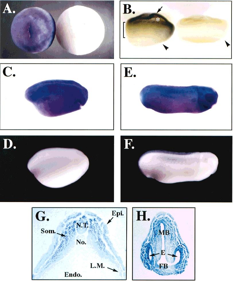
Fig. 4. Temporal and spatial expression of CNBP mRNA during Xenopus early embryogenesis. A-F: Whole mount in situ localization of XCNBP. G,H: Transverse sections of C and E, respectively. A,B: Early gastrula stage (10.25-10.5). These embryos are viewed from the animal pole and side, respectively. Dorsal is to the right. The asterisk indicates the blastocoele cavity, the arrowheads indicate the dorsal lip of the blastopore, and the arrow indicates XCNBP expression in the animal cap region (above the blastocoele cavity). XCNBP staining in the marginal zone is shown by the bracket. In A and B, the embryo on the right is a sense strand control. C,E: Late neurula (stage 15) and late tailbud (stage 27), respectively (antisense probe). D,F: Late neurula (stage 15) and late tailbud (stage 27), respectively (sense probe). In C-F, dorsal is up and anterior is to the right. Epi., epidermis; Endo., endoderm; E, eyes; FB, forebrain; L.M., lateral mesoderm; MB, midbrain; N.T., neural tube; No., notochord; Som., somitic mesoderm.
Image published in: Flink IL et al. (1998)
Copyright © 1998. Image reproduced with permission of the Publisher, John Wiley & Sons.
| Gene | Synonyms | Species | Stage(s) | Tissue |
|---|---|---|---|---|
| cnbp.L | cnbp1, dm2, promm, rnf163, xcnbp, zcchc22, znf9 | X. laevis | Throughout NF stage 10.25 to NF stage 10.5 | animal cap marginal zone |
| cnbp.L | cnbp1, dm2, promm, rnf163, xcnbp, zcchc22, znf9 | X. laevis | Throughout NF stage 15 | dorsal neural tube epidermis presomitic mesoderm lateral plate mesoderm |
| cnbp.L | cnbp1, dm2, promm, rnf163, xcnbp, zcchc22, znf9 | X. laevis | Throughout NF stage 27 | midbrain forebrain eye |
Image source: Published
Permanent Image Page
Printer Friendly View
XB-IMG-75705