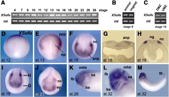
Fig. 2. Gene expression of XSufu in Xenopus embryos. (AâC) RT-PCR of whole embryos (A) and embryonic explants (B,C). Histone H4 was used as RNA loading control. DMZ, dorsal marginal zone; VMZ, ventral marginal zone. (DâM) Whole-mount in situ hybridization of embryos shown in dorsal (D,E), anterior (F,I,J), and lateral views (KâM). Panels (G) and (H) are transversal sections of embryo in (I). anp, anterior neural plate; ba, branchial arch; ea, ear; ey, eye; fb, forebrain; mhb, midâhindbrain boundary; nc, neural crest; ng, neural groove; np, neural plate; ppp, panplacodal primordium, tb, tail bud.
Image published in: Min TH et al. (2011)
Copyright © 2011. Image reproduced with permission of the Publisher, Elsevier B. V.
| Gene | Synonyms | Species | Stage(s) | Tissue |
|---|---|---|---|---|
| sufu.L | sufuh, sufuxl, Xsufu | X. laevis | Sometime during NF stage 12 to NF stage 13 | ectoderm neuroectoderm anterior placodal area |
| sufu.L | sufuh, sufuxl, Xsufu | X. laevis | Sometime during NF stage 15 to NF stage 18 | neural crest neuroectoderm neural groove neural plate neural plate border |
| sufu.L | sufuh, sufuxl, Xsufu | X. laevis | Throughout NF stage 23 | forebrain neural crest eye primordium brain neural tube cranial neural crest |
| sufu.L | sufuh, sufuxl, Xsufu | X. laevis | Throughout NF stage 26 | forebrain neural crest brain eye midbrain-hindbrain boundary otic region pharyngeal arch mandibular arch hyoid arch branchial arch |
| sufu.L | sufuh, sufuxl, Xsufu | X. laevis | Throughout NF stage 32 | forebrain neural crest tail bud brain eye midbrain-hindbrain boundary otic vesicle anterior branchial crest |
Image source: Published
Permanent Image Page
Printer Friendly View
XB-IMG-75915