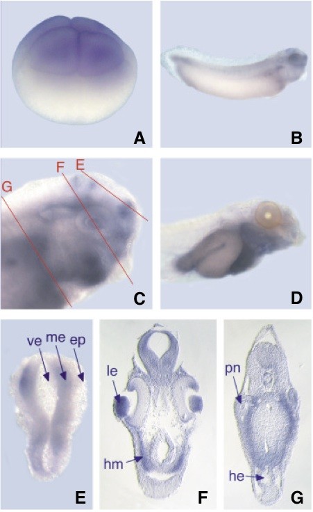
Fig. 6. Whole mount in situ hybridisation of xlFoxP1. (A) 8-cell stage, lateral view; (B) stage 31; (C) stage 35, red lines in (C) indicate the plane of sections shown in (E-G); (D) stage 41, (B-D) are lateral views; (E) horizontal section, anterior is to the bottom; (F,G) transverse sections, dorsal is on top. ep, epidermis; he, heart; hm, head mesenchyme; le, lens; me, mesencephalon; pn, pronephros; ve, brain ventricle.
Image published in: Pohl BS et al. (2005)
Copyright © 2005. Image reproduced with permission of the Publisher.
| Gene | Synonyms | Species | Stage(s) | Tissue |
|---|---|---|---|---|
| foxp1.S | 12cc4, fkh1b, hspc215, qrf1, xlfoxp1 | X. laevis | Throughout NF stage 31 | brain spinal cord lateral plate mesoderm hindbrain forebrain presomitic mesoderm otic vesicle lens placode tail bud proctodeum |
| foxp1.S | 12cc4, fkh1b, hspc215, qrf1, xlfoxp1 | X. laevis | Throughout NF stage 35 and 36 | brain forebrain hindbrain eye lens ciliary marginal zone midbrain roof plate ventricular zone head region mesenchyme foregut trigeminal ganglion |
| foxp1.S | 12cc4, fkh1b, hspc215, qrf1, xlfoxp1 | X. laevis | Throughout NF stage 41 | mesoderm head region mesenchyme endoderm alimentary system stomach intestine foregut |
| foxp1.S | 12cc4, fkh1b, hspc215, qrf1, xlfoxp1 | X. laevis | Throughout NF stage 4 (8-cell) | animal blastomere |
Image source: Published
Permanent Image Page
Printer Friendly View
XB-IMG-76115