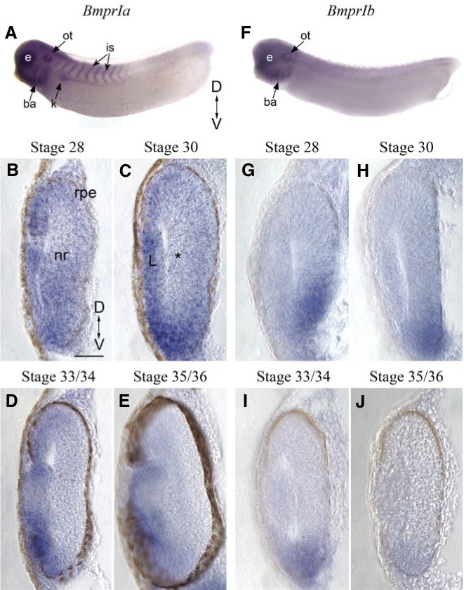
Fig. 3. Distribution of type I Bmp receptor transcripts in the developing Xenopus eye. (A) Wholemount in situ hybridization of a stage 28 embryo using a BmprIa antisense probe. (B-E) Transverse sections through the retinas of Xenopus embryos processed for BmprIa in situ hybridization, at stages 28 (B), 30 (C), 33/34 (D) and 35/36 (E). * in (C) is the forming RGCL. (F) Bmpr1b wholemount in situ hybridization of a stage 30 embryo. (G-J) Transverse retinal sections through stage 28 (G), 30 (H), 33/34 (I) and 35/36 (J) embryos show expression of BmprIb. Scale bar in (B) is 50 μm for (B-E) and (G-J). Abbreviations: D, dorsal; V, ventral; ba, branchial arches; e, eye; k, kidney; is, intersomitic region; L, lens; nr, neural retina; ot, otic vesicle; rpe, retinal pigment epithelium.
Image published in: Hocking JC and McFarlane S (2007)
Copyright © 2007. Image reproduced with permission of the Publisher.
| Gene | Synonyms | Species | Stage(s) | Tissue |
|---|---|---|---|---|
| bmpr1a.S | acvrlk3, alk-3, alk3, cd292, skr5 | X. laevis | Sometime during NF stage 28 to NF stage 29 and 30 | retina eye eye primordium otic vesicle kidney early distal tubule early proximal tubule intersomitic region tail bud brain midbrain hindbrain anterior neural tube pharyngeal arch mandibular arch hyoid arch branchial arch retinal inner nuclear layer retinal outer nuclear layer lens placode retinal ganglion cell layer |
| bmpr1b.L | alk-6, alk6 | X. laevis | Sometime during NF stage 28 to NF stage 29 and 30 | eye ventral retina retinal neural layer retinal ganglion cell layer otic vesicle notochord |
| bmpr1a.S | acvrlk3, alk-3, alk3, cd292, skr5 | X. laevis | Sometime during NF stage 33 and 34 to NF stage 35 and 36 | retina eye eye primordium optic vesicle lens placode ciliary marginal zone |
Image source: Published
Permanent Image Page
Printer Friendly View
XB-IMG-77296