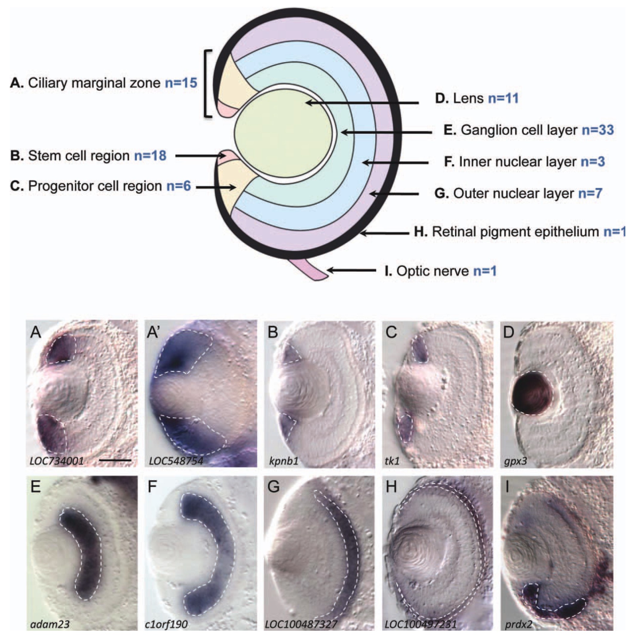
Figure 2 Classification of specific retinal markers retrieved from the screen. Schematic diagram illustrates the chosen subdivisions of the tadpole retina. n indicates the number of markers retrieved from the screen that exhibit specific expression in only one of these compartments. Pictures below are stage 39 transverse retinal sections (dorsal up) that provide typical examples of such markers, with a correspondence to the schematic lettering (A). A fuzzy staining in the CMZ that invades the nuclear layers (A0) is not considered CMZ-specific. White dotted lines delineate the staining. Gene symbol is indicated under each panel. Scale bar = 50 um. Color figure can be viewed in the online issue, which is available at wileyonlinelibrary.com. This material is reproduced with permission of John Wiley & Sons, Inc.
Image published in: Parain K et al. (2012)
Copyright © 2012. Image reproduced with permission of the Publisher, John Wiley & Sons.
| Gene | Synonyms | Species | Stage(s) | Tissue |
|---|---|---|---|---|
| ptmap12 | LOC728026, LOC734001 | X. tropicalis | Throughout NF stage 39 | ciliary marginal zone |
| ddx39a | bat1, bat1l, ddx39, ddxl | X. tropicalis | Throughout NF stage 39 | ciliary marginal zone |
| kpnb1 | imb1, impnb, importin beta, ipo1, ipob, LOC108700788, ntf97 | X. tropicalis | Throughout NF stage 39 | ciliary marginal zone |
| tk1 | X. tropicalis | Throughout NF stage 39 | ciliary marginal zone | |
| gpx3 | gpx3-a, gpx3-b, gpx-p, gshpx-3, gshpx-p | X. tropicalis | Throughout NF stage 39 | lens |
| adam23 | LOC108701467, LOC108702289 | X. tropicalis | Throughout NF stage 39 | retinal ganglion cell layer |
| lurap1 | c1orf190 | X. tropicalis | Throughout NF stage 39 | retinal inner nuclear layer |
| usp21 | LOC100487327, LOC108699609 | X. tropicalis | Throughout NF stage 39 | retinal outer nuclear layer |
| tmem114 | cldn26, LOC100497281 | X. tropicalis | Throughout NF stage 39 | retinal pigmented epithelium |
| prdx2 | nkefb, peroxiredoxin 2, prx-2, prx2, prxii, tdpx1, tpx1 | X. tropicalis | Throughout NF stage 39 | optic nerve |
Image source: Published
Permanent Image Page
Printer Friendly View
XB-IMG-79192