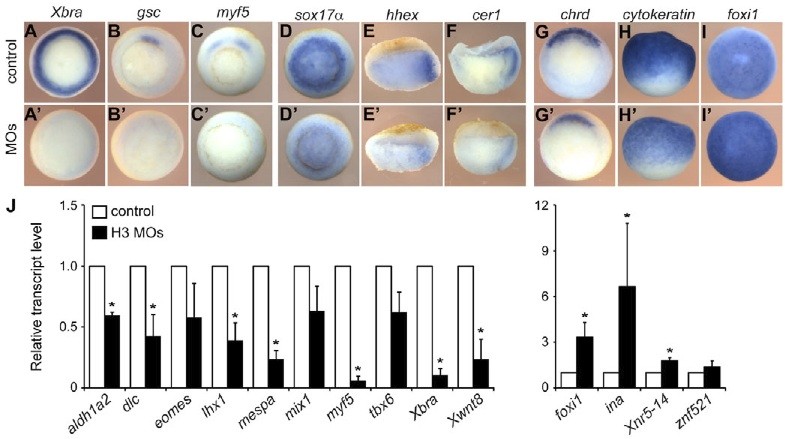
Fig. 2. H3.3/H3-depleted Xenopus embryos fail to express mesodermal marker genes. (A-Iâ²) Control and injected embryos fixed at early gastrula were subjected to RNA in situ hybridization for the analysis of mesodermal (A-Câ²), endodermal (D-Fâ²) and ectodermal (G-Iâ²) marker gene expression. Representative embryos from three experiments are shown. (A-D,G) Vegetal views; (E,F) lateral views of bisected embryos, dorsal towards the right; (H) lateral view; (I) animal view. (J) Expression of selected genes in control and H3 MO-injected embryos at stage 10.5 was measured by qRT-PCR. All values were normalized to ornithine decarboxylase (ODC) and plotted relative to the respective transcript levels in control embryos. Error bars indicate s.d. of three independent experiments. *P<0.05 using a two-tailed Student t-test.
Image published in: Lim CY et al. (2013)
Copyright © 2013. Image reproduced with permission of the Publisher.
| Gene | Synonyms | Species | Stage(s) | Tissue |
|---|---|---|---|---|
| tbxt.S | bra, brachyury, ntl, t, t-a, t-b, X-bra, Xbra, Xbrachyury | X. laevis | Throughout NF stage 10 | mesoderm dorsal marginal zone |
| gsc.L | goosecoid, gsc-a, gsc-b, Xgsc | X. laevis | Throughout NF stage 10 | mesoderm dorsal marginal zone |
| myf5.L | myf-5, Xmyf-5, Xmyf5 | X. laevis | Throughout NF stage 10 | mesoderm marginal zone dorso-lateral marginal zone |
| sox17a.L | sox17 alpha, sox17-alpha, Sox17alpha, tSox17alpha, xsox17, xsox17a, Xsox17-alpha, Xsox17alpha, xSox17alpha1, xSox17alpha2 | X. laevis | Throughout NF stage 10 | endoderm vegetal endoderm blastopore |
| hhex.L | hex, tHex, XHex | X. laevis | Throughout NF stage 10 | endoderm endomesoderm |
| cer1.S | cer, cer-1, Cerberus, dand4, LOC108713989, tCerberus, xcer, xcer-1, xcer1 | X. laevis | Throughout NF stage 10 | endomesoderm endoderm |
| chrd.S | chd, chordin, chrd.1, LOC108716939, X-chordin | X. laevis | Throughout NF stage 10 | dorsal marginal zone ectoderm |
| krt12.4.L | ck81, cytok, cytokeratin, E-keratin, epidermal keratin, Epidermis-specific keratin, EpiK, epiker, epi-keratin, epK, EpK, K81, xK81, xk81a, xk81a1 | X. laevis | Throughout NF stage 10 | ectoderm epidermis animal hemisphere |
| foxi1.L | ectodermally-expressed mesendoderm antagonist, ema, FoxI1e, foxi1e, foxi3, Xema, xfoxi1 | X. laevis | Throughout NF stage 10 | epidermis animal hemisphere ectoderm |
Image source: Published
Permanent Image Page
Printer Friendly View
XB-IMG-79701