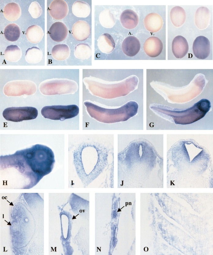
Fig. 2. Localization of InsR transcripts during embryogenesis by whole mount in situ hybridization. (A±G) Expression of InsR mRNA at different stages of embryogenesis (A, stage 61/2; B, stage 9; C, stage 101/2; D, stages 13 (left) and 17 (right); E, stages 23 (left) and 25 (right); F, stage 32; G, stage 37). Control: embryos in the top row of each picture are hybridized with InsR sense probe. Four different views are presented in pictures (A±C): animal (A) or vegetative (V) hemisphere, lateral view (l) or parasagittal section. InsR expression in the anterior part (E) of a stage 37 embryo observed at higher magni®cation. Transverse sections (stage 37) through the encephalon (I, proencephalon; J and K, rhombencephalon), the eye (L: oc, optic cup; l, lens), the otic vesicle (ov, picture M), the pronephros (pn, picture N) and the somites (picture O) readily show that InsR is expressed in these tissues.
Image published in: Groigno L et al. (1999)
Copyright © 1999. Image reproduced with permission of the Publisher, Elsevier B. V.
| Gene | Synonyms | Species | Stage(s) | Tissue |
|---|---|---|---|---|
| insr.L | cd220, hhf5, insulin receptor, Xe-InsR, XTK1, XTK1a, XTK1b | X. laevis | Throughout NF stage 10.5 | animal animal cap |
| insr.L | cd220, hhf5, insulin receptor, Xe-InsR, XTK1, XTK1a, XTK1b | X. laevis | Throughout NF stage 13 | dorsal ectoderm neuroectoderm |
| insr.L | cd220, hhf5, insulin receptor, Xe-InsR, XTK1, XTK1a, XTK1b | X. laevis | Throughout NF stage 23 to NF stage 25 | head region |
| insr.L | cd220, hhf5, insulin receptor, Xe-InsR, XTK1, XTK1a, XTK1b | X. laevis | Throughout NF stage 32 | somite head mesoderm otic vesicle |
| insr.L | cd220, hhf5, insulin receptor, Xe-InsR, XTK1, XTK1a, XTK1b | X. laevis | Throughout NF stage 37 and 38 | somite pronephric kidney pronephric nephron late proximal tubule early distal tubule early proximal tubule pharyngeal arch otic vesicle head region neural tube brain forebrain hindbrain eye retina |
| insr.L | cd220, hhf5, insulin receptor, Xe-InsR, XTK1, XTK1a, XTK1b | X. laevis | Sometime during NF stage 6.5 to NF stage 9 | animal cap animal |
Image source: Published
Permanent Image Page
Printer Friendly View
XB-IMG-79711