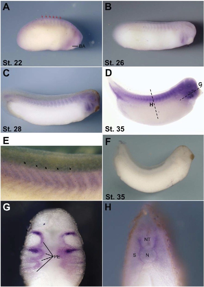
Fig. 5. Spatial expression of pax9 transcripts during Xenopus development. Whole-mount pax9 in situ hybridization. A: Lateral view, anterior is to the right. A: At stage 22, pax9 is weakly expressed in the branchial arches and in the somite (red arrows). B: pax9 expression is increased and its localization advances progressively to caudal region in the stage 26 (B), stage 28 (C), and stage 35 embryos (D). E: Magnification of the dorsal region of C at the somites level. Black arrows indicate the intersomitic space. F: Whole-mount in situ hybridization with pax9 sense probe. G,H: Transverse sections of stage 35 embryos at the branchial arches level (G) and trunk (H).
Image published in: Sánchez RS and Sánchez SS (2013)
Copyright © 2013. Image reproduced with permission of the Publisher.
| Gene | Synonyms | Species | Stage(s) | Tissue |
|---|---|---|---|---|
| pax9.L | Pax-9, sthag3, xpax-9, xpax9 | X. laevis | Throughout NF stage 22 | somite pharyngeal endoderm |
| pax9.L | Pax-9, sthag3, xpax-9, xpax9 | X. laevis | Throughout NF stage 26 | somite pharyngeal endoderm |
| pax9.L | Pax-9, sthag3, xpax-9, xpax9 | X. laevis | Throughout NF stage 28 | somite pharyngeal endoderm |
| pax9.L | Pax-9, sthag3, xpax-9, xpax9 | X. laevis | Throughout NF stage 35 and 36 | pharyngeal endoderm somite sclerotome otic vesicle head region pharyngeal pouch |
Image source: Published
Permanent Image Page
Printer Friendly View
XB-IMG-81529