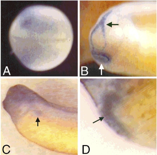
Fig. 5. Xmad4 expression profile by whole-mount in situ hybridization. In all panels, anterior is to the left. A: Dorsal view of a late neurula embryo showing strong Xmad4 expression in the ectoderm immediately adjacent to the neural tube. B: Anterior lateral view of an early tailbud stage embryo. Note expression in the Y-shaped hatching gland (black arrow) and the more ventral cement gland (white arrow). C: Lateral view of a mid-tailbud stage embryo showing Xmad4 expression in the pronephros (black arrow). The darkness of the head is background, due to the extended period of staining required to visualize the relatively low level of transcription in the kidney. D: Close-up view of a mid tailbud stage embryo. Xmad4 expression is detected in the embryonic liver (arrow).
Image published in: Newman CS and Krieg PA (1999)
Copyright © 1999. Image reproduced with permission of the Publisher, John Wiley & Sons.
| Gene | Synonyms | Species | Stage(s) | Tissue |
|---|---|---|---|---|
| mxd4.S | bhlhc12, mad4, mst149, mstp149, mxd4-a, mxd4-b, xmad4 | X. laevis | Throughout NF stage 16 | ectoderm lateral plate mesoderm |
| mxd4.S | bhlhc12, mad4, mst149, mstp149, mxd4-a, mxd4-b, xmad4 | X. laevis | Throughout NF stage 24 | cement gland hatching gland |
| mxd4.S | bhlhc12, mad4, mst149, mstp149, mxd4-a, mxd4-b, xmad4 | X. laevis | Throughout NF stage 29 and 30 | liver pronephric kidney head region liver primordium |
Image source: Published
Permanent Image Page
Printer Friendly View
XB-IMG-81763