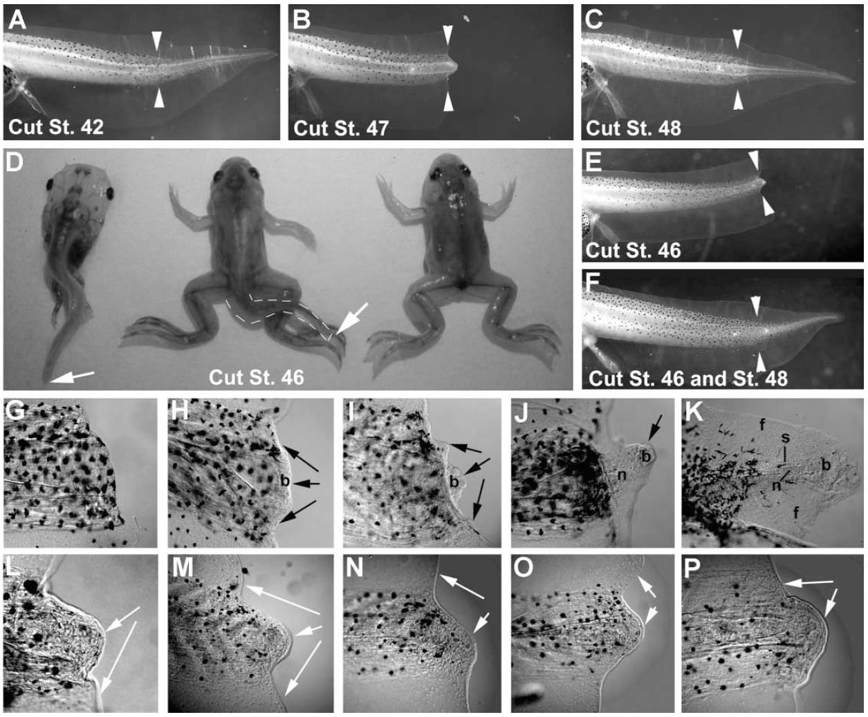
Figure 1. Axial Regenerative Ability in Xenopus laevis Tadpoles Is Lost at Stage 45/47 and Regained at Stage 48 and Later. White arrowheads show the level of tail amputation where appropriate. (A-C) Tadpoles were subjected to removal of 50% of the postanal tail at different stages of development and allowed to recover for 7 days. Stage 42 (A), stage 47 (B), and stage 48 (C).(D) Tadpoles subjected to 50% tail removal at stage 46 do not regenerate a tail, but develop normally, and the remaining stump is resorbed normally during metamorphosis. Left to right: stage 58; stage 63; stage 66.(E and F) Tadpole tails amputated at stage 46 (E) will regenerate if they are cut again after stage 48 (F). (G-P) Tail stumps partially cleared in glycerol and viewed under Nomarski optics. (G-K) Regeneration process in stage 49 tadpoles. Wound epidermis is indicated by black arrows. (G) Stage 49 tail fixed immediately after amputation. (H) After 24 hr, the wound epithelium has formed and blastemal cells are appearing. (I) Within 48 hr, blastemal cells have migrated into the center of the lesion and are proliferating ('cone 'stage). (J) After 3 days, the proximal blastemal cells are beginning to redifferentiate into the constituent tissues of the tail. (K) By 4-5 days, new notochord, spinal cord, fin, and presomitic mesoderm have formed and melanocytes (black pigment cells) are migrating into the new tail. (L-P) In contrast, tails cut at stage 47 do not regenerate or form a blastema, and the cut surface becomes covered with a skin-like epithelium (white arrows). (L) By 24 hr postamputation, a skin-like epithelium has formed over the cut surface of the tail stump (white arrows). The tissue behind this epithelium does not form a blastema. (M-P) The wound area does not alter over time, and there is no regeneration of tail tissues. (M-P) After 48 hr (M), 3 days (N), 4 days (O), and 7 days (P) postamputation. All tail stumps are lateral views oriented anterior to the left, and dorsal uppermost. b, regeneration bud; f, fin; n, notochord; s, spinal cord.
Image published in: Beck CW et al. (2003)
Copyright © 2003. Image reproduced with permission of the Publisher, Elsevier B. V.
Permanent Image Page
Printer Friendly View
XB-IMG-82781